Japanese
English
症例
脳有鉤嚢虫症の1手術例
Cerebral Cysticercosis: Report of an operated case
清水 幸彦
1,2
,
香川 茂樹
1,2
,
小沼 武英
1
Yukihiko SHIMIZU
1,2
,
Shigeki KAGAWA
1,2
,
Takehide ONUMA
1
1仙台市立病院脳神経外科
2現籍 東北大学脳研脳神経外科
1Department of Neurosurgery, Sendai City Hospital
キーワード:
Cerebral cysticercosis
,
CT
Keyword:
Cerebral cysticercosis
,
CT
pp.209-213
発行日 1986年2月10日
Published Date 1986/2/10
DOI https://doi.org/10.11477/mf.1436202152
- 有料閲覧
PPV購入案内
1,320円 (1,200円+税10%) こちらは、209ページから213ページの文献です。
PPV(ペイ・パー・ビュー)とは、論文(記事)単位で購入・閲覧ができるサービスです。
購入には医書.jp本会員登録が必要です。
会員登録完了後に改めて上記「購入サイトへ」を選択してください。
または「購入サイトへ」選択後に「購入ログイン」、「新規会員登録」の順に進んでください。
会員登録について詳しくはをご確認ください。
購入には医書.jp本会員登録が必要です。
会員登録完了後に改めて上記「購入サイトへ」を選択してください。
または「購入サイトへ」選択後に「購入ログイン」、「新規会員登録」の順に進んでください。
会員登録について詳しくはをご確認ください。
- Abstract 文献概要
- 1ページ目 Look Inside
I.はじめに
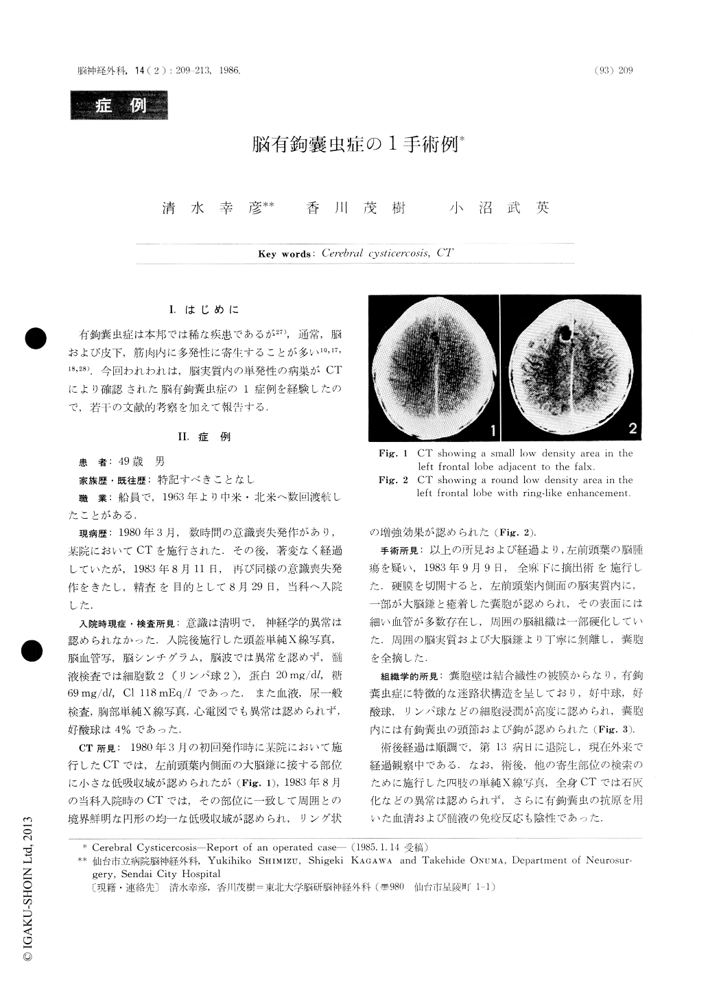
有鉤嚢虫症は本邦では稀な疾患であるが27),通常,脳および皮下,筋肉内に多発性に寄生することが多い10,17,18,28).今回われわれは,脳実質内の単発性の病巣がCTにより確認された脳有鉤嚢虫症の1症例を経験したので,若干の文献的考察を加えて報告する.
A rare case of cerebral cysticercosis is reported. The patient, a 49-year-old man, had been to Central America several times since 1963. He had an episode of seizure in March 1980 and again in August 1983, and was admitted to our hospital.
Skull X-ray, cerebral angiography, scintigraphy, EEG, CSF examination, Chest X-ray, blood and urine examinations were all normal. CT scan only revealed a small lesion in the left frontal lobe as a well cir-cumscribed round low density area with ring-like enhancement.
An operation was performed for a suspected brain tumor.
Copyright © 1986, Igaku-Shoin Ltd. All rights reserved.