Japanese
English
- 有料閲覧
- Abstract 文献概要
- 1ページ目 Look Inside
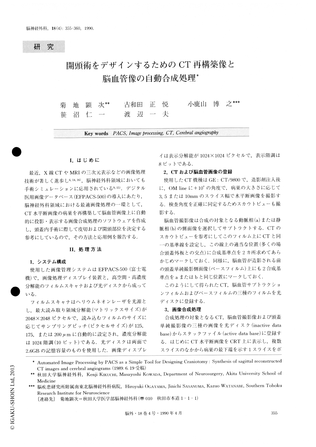
I.はじめに 最近,X線CTやMRIの三次元表示などの画像処理技術が著しく進歩し6,14,16),脳神経外科領域においても手術シミュレーションに応用されている9,15).デジタル医用画像データベース(EFPACS−500)の導入にあたり,脳神経外科領域における最適画像処理の一環として,CT水平断画像の病巣を再構築して脳血管画像上に自動的に投影・表示する画像合成処理のソフトウェアを作成し,頭蓋内手術に際して皮切および開頭部位を決定する参考にしているので,その方法と応用例を報告する.
Abstract
Software has been developed to produce sagittal im-ages of the brain recontracted from axial CT slices and automatically superimpose them on the lateral views of cerebral angiograms. A commercially available system for digitization, processing, display and computer stor-age of film radiographs (EFPACS-500) was used in the present studies. Axial CT scans are obtained in either 3, 5 or 10mm thicknesses parallel to the base line at the 10 degree angle to the OM line, and processed into the digital storage.

Copyright © 1990, Igaku-Shoin Ltd. All rights reserved.


