Japanese
English
- 有料閲覧
- Abstract 文献概要
- 1ページ目 Look Inside
I.はじめに
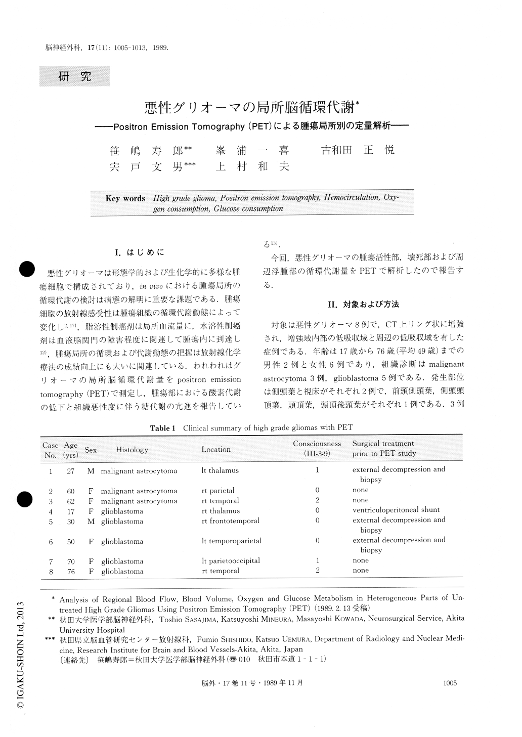
悪性グリオーマは形態学的および生化学的に多様な腫瘍細胞で構成されており,in vivoにおける腫瘍局所の循環代謝の検討は病態の解明に重要な課題である.腫瘍細胞の放射線感受性は腫瘍組織の循環代謝動態によって変化し2,17),脂溶性制癌剤は局所血流量に,水溶性制癌剤は血液脳関門の障害程度に関連して腫瘍内に到達し12),腫瘍局所の循環および代謝動態の把握は放射線化学療法の成績向上にも大いに関連している.われわれはグリオーマの局所脳循環代謝量をpositron emissiontomography(PET)で測定し,腫瘍部における酸素代謝の低下と組織悪性度に伴う糖代謝の亢進を報告している13).
今回,悪性グリオーマの腫瘍活性部,壊死部および周辺浮腫部の循環代謝量をPETで解析したので報告する.
A high resolution positron emission tomography (PET), HEADTOME III, has enabled us to visualize heterogeneous parts, i.e. viable, necrotic, and edematic portions in malignant gliomas, and to quantify regional hemocirculation and metabolism of the tumors using 15O and 18F-fluorodeoxyglucose tracers.
Hemocirculatory and metabolic indices of regional cerebral blood flow (rCBF), blood volume (rCBV), oxygen extraction fraction (rOEF), oxygen consumption (rCMRO2) and glucose consumption (rCMRGl) were studied in eight patients with untreated malignant gliomas.

Copyright © 1989, Igaku-Shoin Ltd. All rights reserved.


