Japanese
English
- 有料閲覧
- Abstract 文献概要
- 1ページ目 Look Inside
I.はじめに
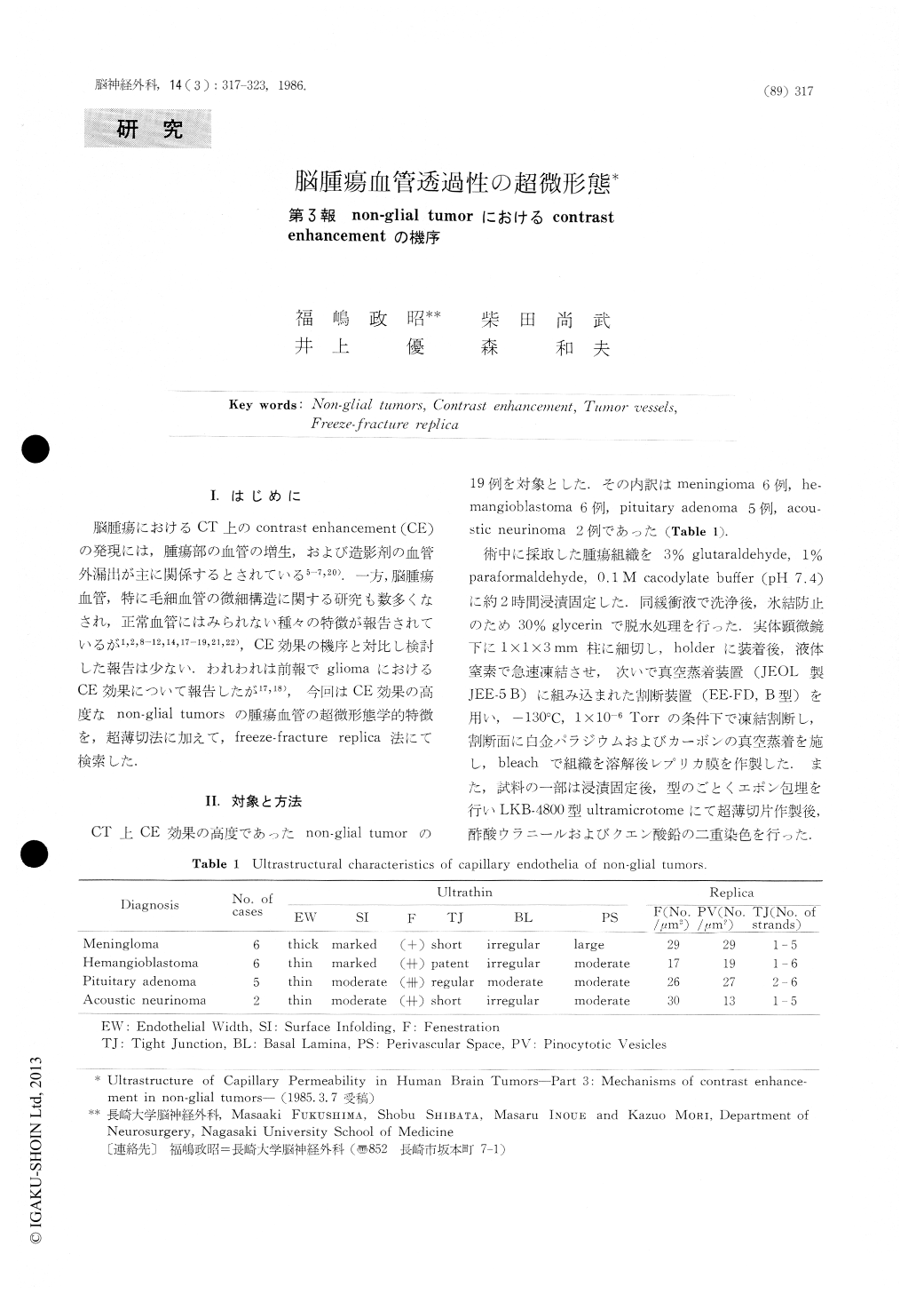
脳腫瘍におけるCT上のcontrast enhancement(CE)の発現には,腫瘍部の血管の増生,および造影剤の血管外漏出が主に関係するとされている5-7,20).一方,脳腫瘍血管,特に毛細血管の微細構造に関する研究も数多くなされ,正常血管にはみられない種々の特徴が報告されているが1,2,8-12,14,17-19,21,22),CE効果の機序と対比し検討した報告は少ない.われわれは前報でgliomaにおけるCE効果について報告したが17,18),今回はCE効果の高度なnon-glial tumorsの腫瘍血管の超微形態学的特徴を,超薄切法に加えて,freeze-fracture replica法にて検索した.
In order to elucidate mechanisms of contrast en-hancement on computed tomography observed in non-glial tumors, tumors vessels were studied with con-ventional ultrathin section and freeze-fracture replica techniques.
The materials were obtained from surgically re-moved specimens in 19 cases of tumors (6 of menin-gioma, 6 of hemangioblastoma, 5 of pituitary adenoma, and 2 of acoustic neurinoma).

Copyright © 1986, Igaku-Shoin Ltd. All rights reserved.


