Japanese
English
- 有料閲覧
- Abstract 文献概要
- 1ページ目 Look Inside
I.はじめに
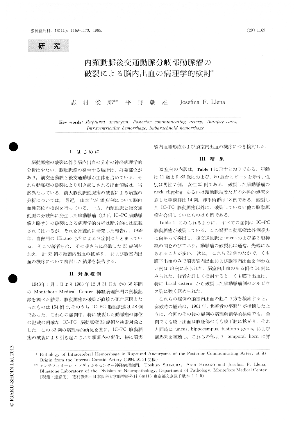
脳動脈瘤の破裂に伴う脳内出血の分布の神経病理学的分析は少ない.脳動脈瘤の発生する場所は,好発部位があり,前交通動脈と後交通動脈が主体を占めている.それら動脈瘤の破裂により引き起こされる出血領域は,当然異なっている.前大脳動脈動脈瘤の破裂による病態の分析については,最近,山本15)が48症例について脳内血腫部位の検討を行つている.一方,内頸動脈と後交通動脈の分岐部に発生した脳動脈瘤(以下,IC-PC脳動脈瘤と略す)の破裂による病理学的分析は断片的には記載されてはいるが,それを系統的に研究した報告は,1959年,当部門のHiranoら6)による9症例にとどまっている.そこで著者らは,その後さらに経験した23症例を加え,計32例の頭蓋内出血の拡がり,および脳室内出血の機序について検討した結果を報告する.
There were thirty-two autopsied cases of ruptured aneurysms at the junction of the internal carotid and posterior communicating arteries in the file of Montefiore Medical Center from 1948 to 1983 (Table 1).
The age range of the patients was 11-83 years. Seven were men and twenty-five were women. Fifteen had previous surgery; either clipping of the neck of the aneurysm or ligation of the common carotid artery.
Analysis of the hemorrhage associated with the ruptured aneurysms revealed two distinct patterns.

Copyright © 1985, Igaku-Shoin Ltd. All rights reserved.


