Japanese
English
- 有料閲覧
- Abstract 文献概要
- 1ページ目 Look Inside
I.はじめに
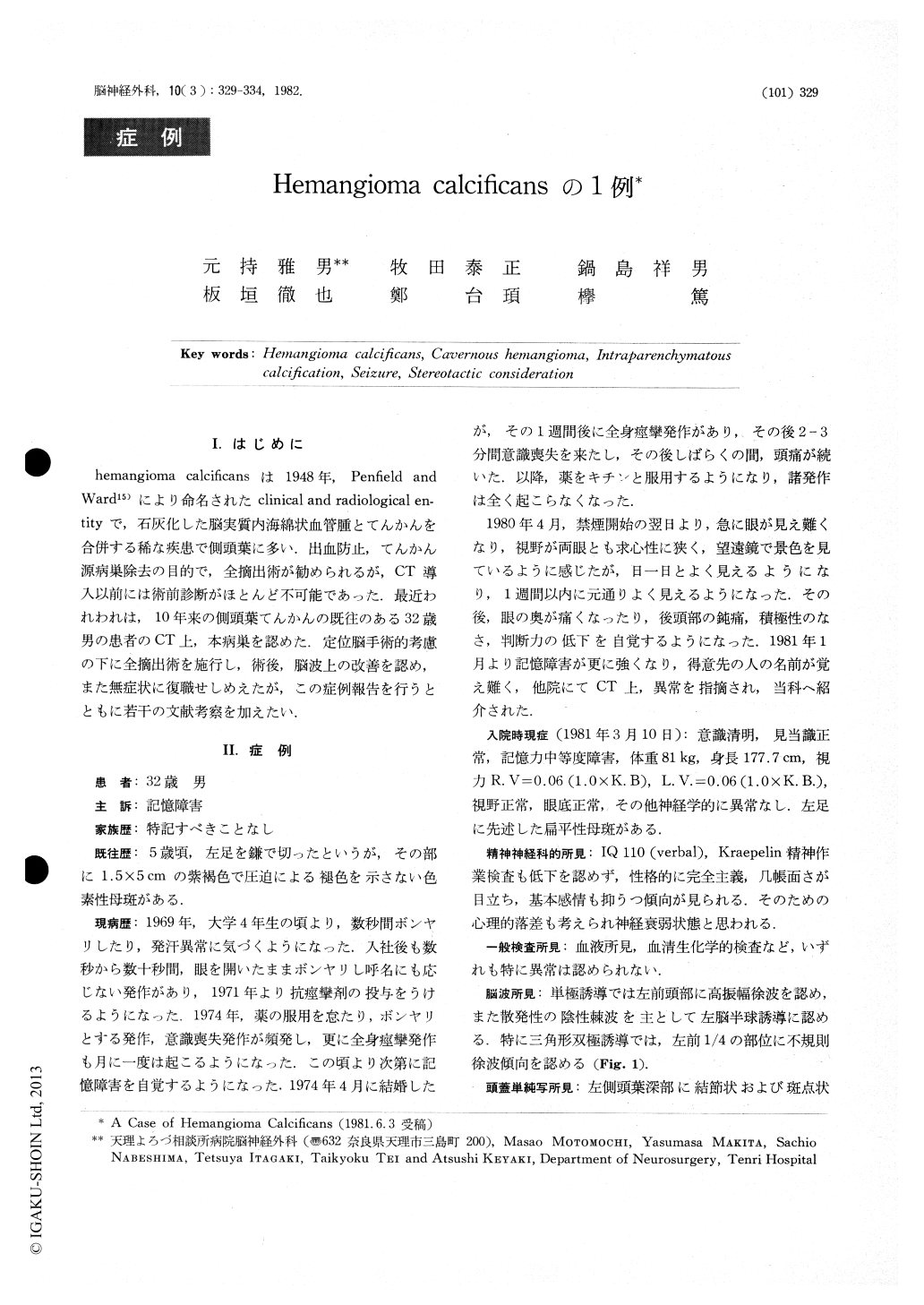
hemangioma calcificansは1948年,Penfield andWard15)により命名されたclinical and radiological en-tityで,石灰化した脳実質内海綿状血管腫とてんかんを合併する稀な疾患で側頭葉に多い.出血防止,てんかん源病巣除去の目的で,全摘出術が勧められるが,CT導入以前には術前診断がほとんど不可能であった.最近われわれは,10年来の側頭葉てんかんの既往のある32歳男の患者のCT上,本病巣を認めた.定位脳手術的考慮の下に全摘出術を施行し,術後,脳波上の改善を認め,また無症状に復職せしめえたが,この症例報告を行うとともに若干の文献考察を加えたい.
A 32-year-old male, who had had temporal lobe seizurefor the past 10 years, was admitted to the neurosurgicalinstitute of Tenri Hospital on March 10, 1981.
Physical examination on admission revealed some memorydisturbance, neuroasthenic tendency and a purplish nevusin the left foot. Plain x-ray series of the skull showedseveral nodular calcified lesions in the medial aspect of theleft temporal lobe. Electroencephalography showedsporadic negative spikes and irregular slow waves dominantin the left anterior quadrant of the head.

Copyright © 1982, Igaku-Shoin Ltd. All rights reserved.


