Japanese
English
症例
Dumbbell形頸部神経鞘腫の椎骨動脈撮影の特徴
Vertebral Angiography of Cervical Dumbbell Neurinomas
佐伯 直勝
1
,
佐藤 章
1
,
北原 聰樹
1
,
辛 秀雄
1
,
山浦 晶
1
,
末吉 貫爾
2
,
牧野 博安
1
Neokatsu SAEKI
1
,
Akira SATO
1
,
Satoki KITAHARA
1
,
Hideo SHIN
1
,
Akira YAMAURA
1
,
Kanji SUEYOSHI
2
,
Hiroyasu MAKINO
1
1千葉大学脳神経外科
2千葉県がんセンター脳神経外科
1Department of Neurosurgery, Chiha University School of Medicine
2Department of Neurosurgery, Chiha Cancer Center Hospital
キーワード:
Spinal tumor
,
Dumbbell tumor Neurinoma Angiography
Keyword:
Spinal tumor
,
Dumbbell tumor Neurinoma Angiography
pp.1287-1294
発行日 1981年10月10日
Published Date 1981/10/10
DOI https://doi.org/10.11477/mf.1436201416
- 有料閲覧
- Abstract 文献概要
- 1ページ目 Look Inside
I.はじめに
頸部脊髄腫瘍の診断には,従来,頸椎単純撮影,myelography,血管撮影などがあり,最近ではspinal CTが登場し,その診断技術は大きく進歩している.
頸部Dumbbell(以下ダンベル)形神経鞘腫の椎骨動脈撮影の重要性は,1952年のLoveに始まり10),以来,いくつかの症例報告により強調されてきた1-5,8,9,13,17).他の診断法と比較した場合,この種の腫瘍における血管撮影の診断価値は,椎骨動脈の閉塞,圧排編位の有無とその方向,腫瘍のvascularityなどの手術時に不可欠な情報を得られることに要約される.
Three cases of cervical dumbbell neurinotnas were reported.
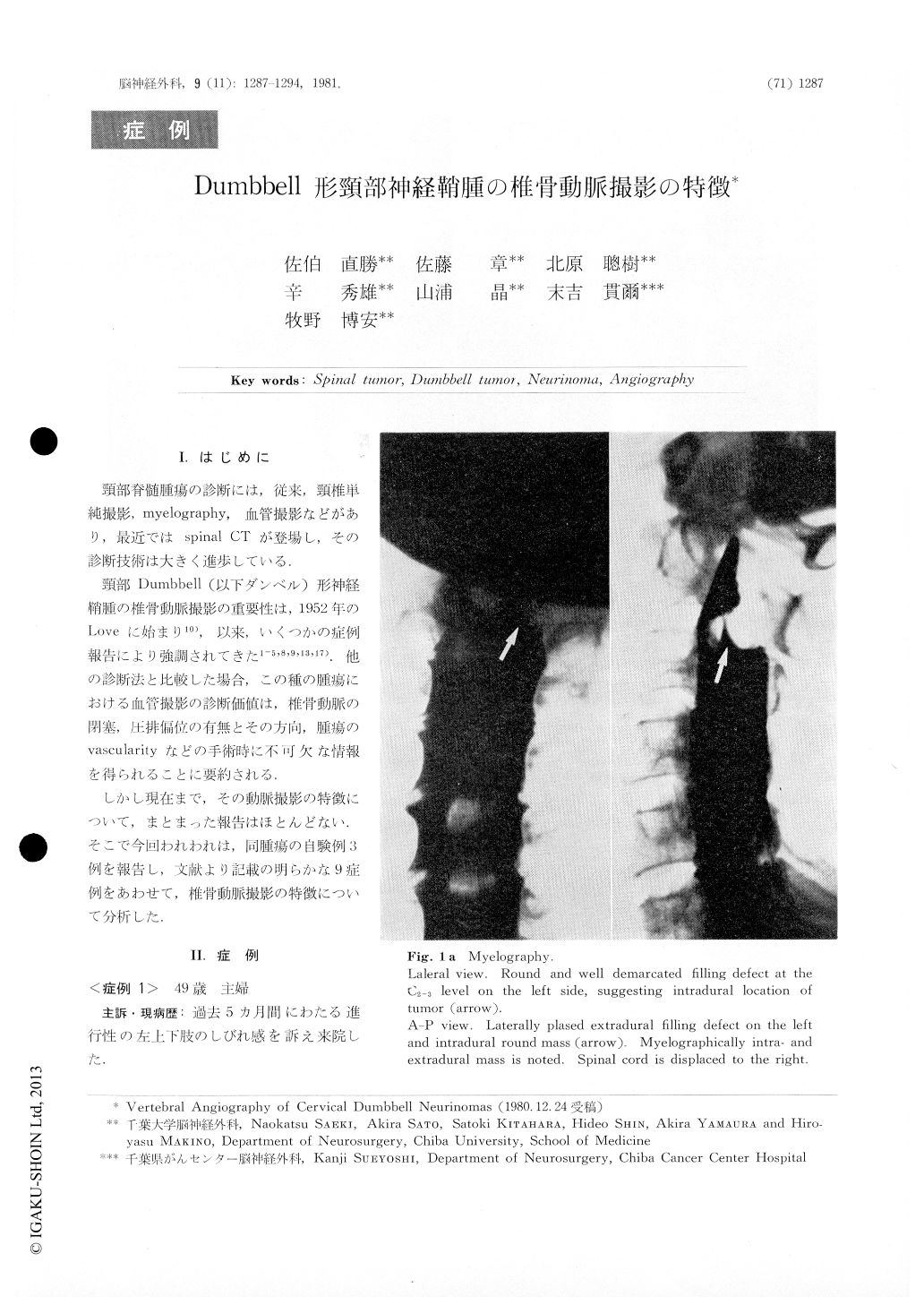
Case. 1. A 49-year-old housewife had left C2-3 intraand extradural neurinoma. The left vertebral angiography showed narrowing and anterior shift of the vertebral artery at C2 level with tumor stain fed by a radicular artery of the same artery.

Copyright © 1981, Igaku-Shoin Ltd. All rights reserved.


