Japanese
English
- 有料閲覧
- Abstract 文献概要
- 1ページ目 Look Inside
- 参考文献 Reference
Ⅰ.はじめに
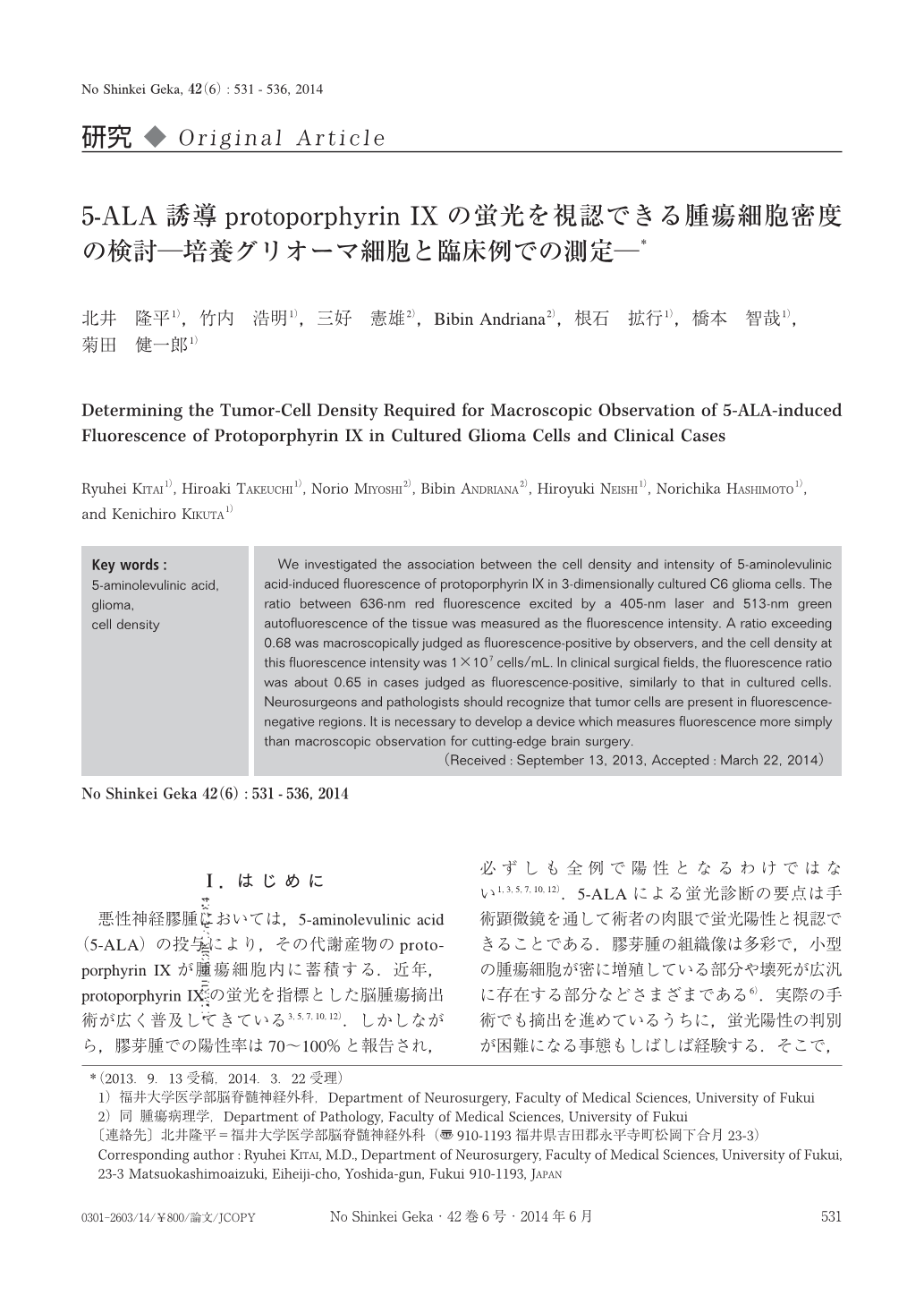
悪性神経膠腫においては,5-aminolevulinic acid(5-ALA)の投与により,その代謝産物のprotoporphyrin Ⅸが腫瘍細胞内に蓄積する.近年,protoporphyrin Ⅸの蛍光を指標とした脳腫瘍摘出術が広く普及してきている3,5,7,10,12).しかしながら,膠芽腫での陽性率は70~100%と報告され,必ずしも全例で陽性となるわけではない1,3,5,7,10,12).5-ALAによる蛍光診断の要点は手術顕微鏡を通して術者の肉眼で蛍光陽性と視認できることである.膠芽腫の組織像は多彩で,小型の腫瘍細胞が密に増殖している部分や壊死が広汎に存在する部分などさまざまである6).実際の手術でも摘出を進めているうちに,蛍光陽性の判別が困難になる事態もしばしば経験する.そこで,蛍光について最も基本的な腫瘍細胞密度に注目し,培養細胞を用いて検討した.研究手法は,培養細胞を腫瘍組織塊に近似させるため,三次元的に分布させ,蛍光の陽性度とスペクトラム測定を行った.さらに,実際の臨床例において組織中protoporphyrin Ⅸ濃度,蛍光スペクトラムを検討し,培養細胞での実験結果と対比した.
We investigated the association between the cell density and intensity of 5-aminolevulinic acid-induced fluorescence of protoporphyrin Ⅸ in 3-dimensionally cultured C6 glioma cells. The ratio between 636-nm red fluorescence excited by a 405-nm laser and 513-nm green autofluorescence of the tissue was measured as the fluorescence intensity. A ratio exceeding 0.68 was macroscopically judged as fluorescence-positive by observers, and the cell density at this fluorescence intensity was 1×10⁷ cells/mL. In clinical surgical fields, the fluorescence ratio was about 0.65 in cases judged as fluorescence-positive, similarly to that in cultured cells. Neurosurgeons and pathologists should recognize that tumor cells are present in fluorescence-negative regions. It is necessary to develop a device which measures fluorescence more simply than macroscopic observation for cutting-edge brain surgery.

Copyright © 2014, Igaku-Shoin Ltd. All rights reserved.


