Japanese
English
- 有料閲覧
- Abstract 文献概要
- 1ページ目 Look Inside
- 参考文献 Reference
Ⅰ.はじめに
2008年7月,山形大学医学部附属病院に1.5テスラ高磁場術中MRIシステムが導入され,脳腫瘍,脳血管障害,てんかんなど種々の脳神経外科手術に用いている.術中MRIシステムの効用は,①残存腫瘍の把握とup-date navigation,②追加切除による悪性脳腫瘍の手術摘出率の上昇,悪性脳腫瘍の治療成績の向上をもたらすことである1,4,11).またさらに③術中の合併症・イベントをいち早くモニタリングすることにより手術の安全性向上にも貢献すると考えられている.しかしこれまで,術中出血などを術中MRIシステムにて検出し得たという少数の報告はあるが10,12),術中画像の撮像条件信号強度について報告したものはない.今回われわれは,右側頭葉悪性神経膠腫摘出術中に対側急性硬膜下血腫を来したものの,術中MRIにて早期に発見し得たことで新たな脱落症状なく治療を行うことができた1例を経験した.本症例ではT1強調,T2強調,FLAIR画像などの他に拡散強調画像,T2*強調画像の撮影を施行しており,今回は超急性期の出血の信号強度とその解釈・診断につき考察を加え報告する.
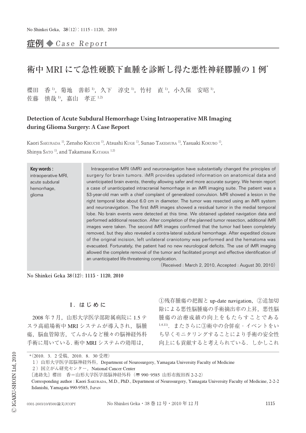
Intraoperative MRI (iMR) and neuronavigation have substantially changed the principles of surgery for brain tumors. iMR provides updated information on anatomical data and unanticipated brain events,thereby allowing safer and more accurate surgery. We herein report a case of unanticipated intracranial hemorrhage in an iMR imaging suite. The patient was a 53-year-old man with a chief complaint of generalized convulsion. MRI showed a lesion in the right temporal lobe about 6.0 cm in diameter. The tumor was resected using an iMR system and neuronavigation. The first iMR images showed a residual tumor in the medial temporal lobe. No brain events were detected at this time. We obtained updated navigation data and performed additional resection. After completion of the planned tumor resection,additional iMR images were taken. The second iMR images confirmed that the tumor had been completely removed,but they also revealed a contra-lateral subdural hemorrhage. After expedited closure of the original incision,left unilateral craniotomy was performed and the hematoma was evacuated. Fortunately,the patient had no new neurological deficits. The use of iMR imaging allowed the complete removal of the tumor and facilitated prompt and effective identification of an unanticipated life-threatening complication.

Copyright © 2010, Igaku-Shoin Ltd. All rights reserved.


