Japanese
English
- 有料閲覧
- Abstract 文献概要
- 1ページ目 Look Inside
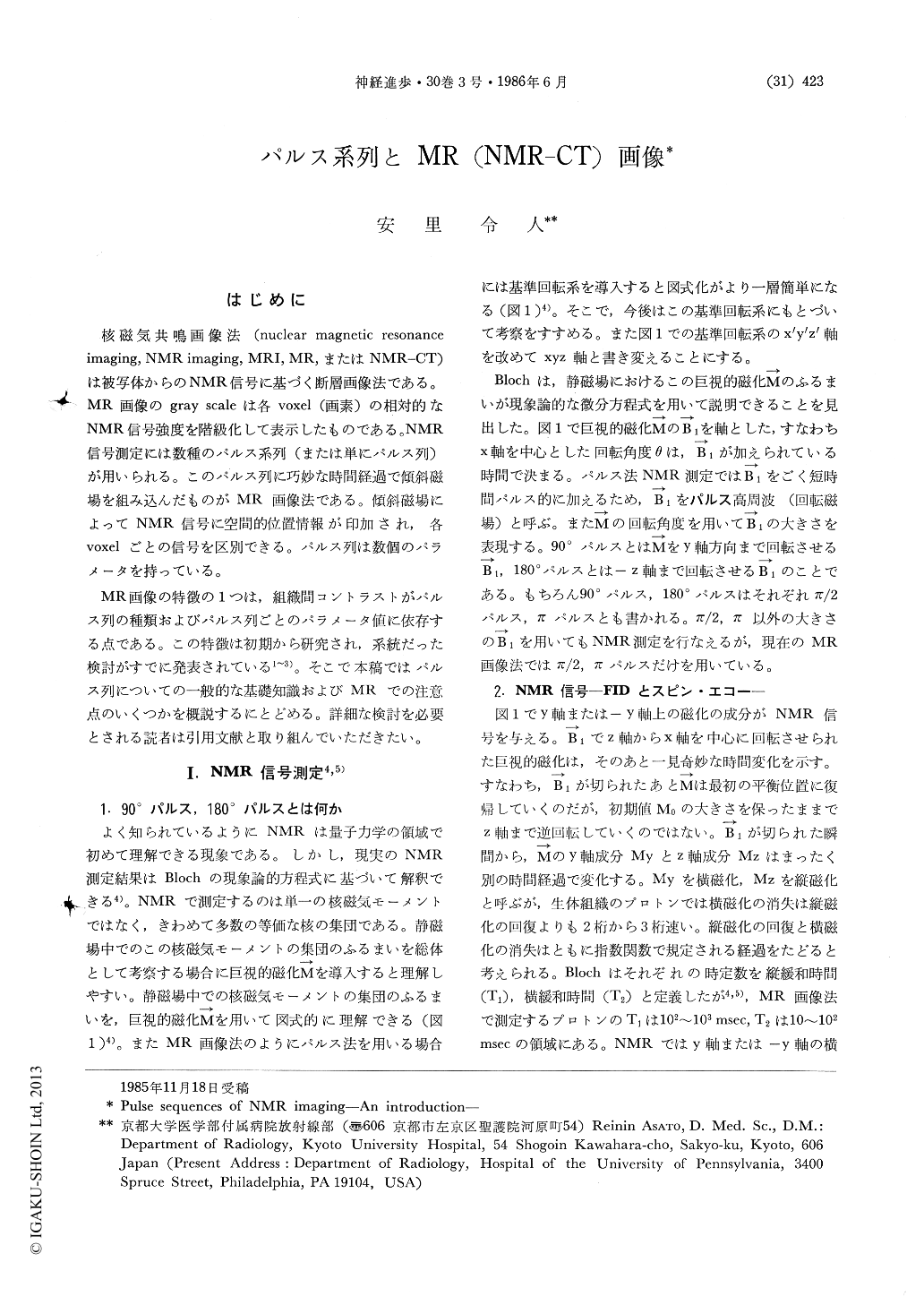
はじめに
核磁気共鳴画像法(nuclear magnetic resonanceimaging,NMR imaging,MRI,MR,またはNMR-CT)は被写体からのNMR信号に基づく断層画像法である。MR画像のgray scalcは各voxel(画素)の相対的なNMR信号強度を階級化して表示したものである。NMR信号測定には数種のパルス系列(または単にパルス列)が用いられる。このパルス列に巧妙な時間経過で傾斜磁場を組み込んだものがMR画像法である。傾斜磁場によってNMR信号に空間的位置情報が印加され,各voxelごとの信号を区別できる。パルス列は数個のパラメータを持っている。
MR画像の特徴の1つは,組織間コントラストがパルス列の種類およびパルス列ごとのパラメータ値に依存する点である。この特徴は初期から研究され,系統だった検討がすでに発表されている1〜3)。そこで本稿ではパルス列についての一般的な基礎知識およびMRでの注意点のいくつかを概説するにとどめる。詳細な検討を必要とされる読者は引用文献と取り組んでいただきたい。
The most prominent characteristic of MR images is variety in contrast. Though in the spin echo (SE) (TR=400 msec/TE=25 msec) image, for example, cerebral gray matter is darker than white matter, in SE (2000/20) image gray matter is shown as the bright area, and white matter apears dark. It has critical importance in MR imaging to select the images whose cotrast is best suited for diagnostic purpose.
We should start to consider the quality of contrast, as high quality images are preferable. Quality of image contrast should be evaluated by the special function of signal to noise ratio and signal intensity ratio between the tissues (Edelstein WA, 1983; Wehrli FW, 1984). However, we can discuss the criteria how to select the images of diagnostically suitable contrast, referring to the well-known signal intensity functions (Young IR, 1982), as far as we consider the effects of imaging parameters on contrast.

Copyright © 1986, Igaku-Shoin Ltd. All rights reserved.


