Japanese
English
- 有料閲覧
- Abstract 文献概要
- 1ページ目 Look Inside
I.はじめに
実験水頭症の研究において,いかにして水頭症を作成するかという点は重要である。現在一般に行なわれているのは,cisterna magnaより異物(カオリン,シリコンオイル)を注入し,頭蓋底槽および,foramen of Magendie,foramen of Luschkaを閉塞させる方法である。そのほかには,中脳水道に,balloonを挿入し,中脳水道閉塞による急性水頭症を作成する方法もある。このほかにもいろいろの方法1)は行なわれて来ているが,現在水頭症の研究といえば,カオリン水頭症がもっとも広く行なわれている力法といえる。しかしこの方法の欠点は,人工的に脳底部くも膜下腔で,カオリンによる炎症性癒着反応を起こさせることであり,とくに小児の先天性水頭症の実験モデルとしては,適当とはいえない。それでは,実験動物において,先天性に水頭症を呈する動動はあるかということになる。mouseで,先天性奇形の一つとして水頭症を呈するものは,hy-1 mice(hydrocephalus-1)Clark2)(1932),hy-2 mice(hydrocephalus-2)Zimmerman3)(1934),hy-3 mice(hydrocephalus-3)Gruneberg4)(1943),chmice(congenital hydrocephalus)Grüneberg5)(1943)などである。
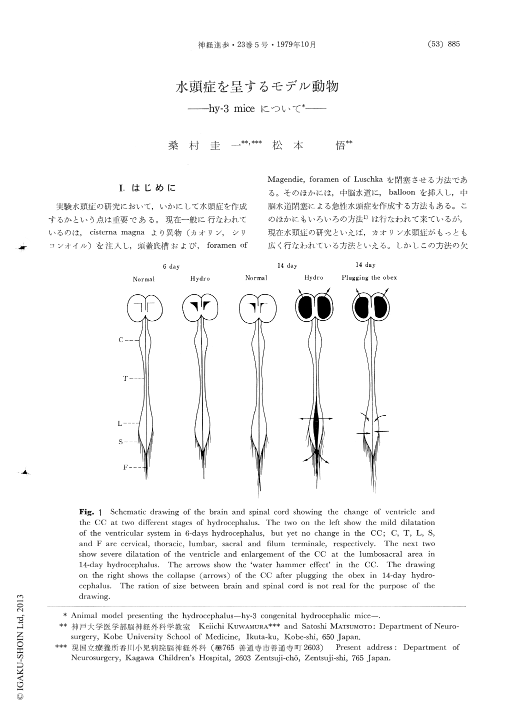
Anatomical and physiological aspects of a murine model of hydrocephalus (hy-3) were reviewed. Especially the brain edema (or interstitial brain edema.), which were found to exist in the white matter at the time of maximum dilatation of the ventricle, were summarized based on the electron microscopic studies. Special attentions were focused on the spinal central canal (CC) of this congenital hydrocephalic mice. And it's histological and physiological studies disclosed the following findings: (1) Dilatation of the CCin hydrocephalic mice with communication between the ventricular system and the CC through the obex.

Copyright © 1979, Igaku-Shoin Ltd. All rights reserved.


