Japanese
English
- 有料閲覧
- Abstract 文献概要
- 1ページ目 Look Inside
Ⅰ.緒言
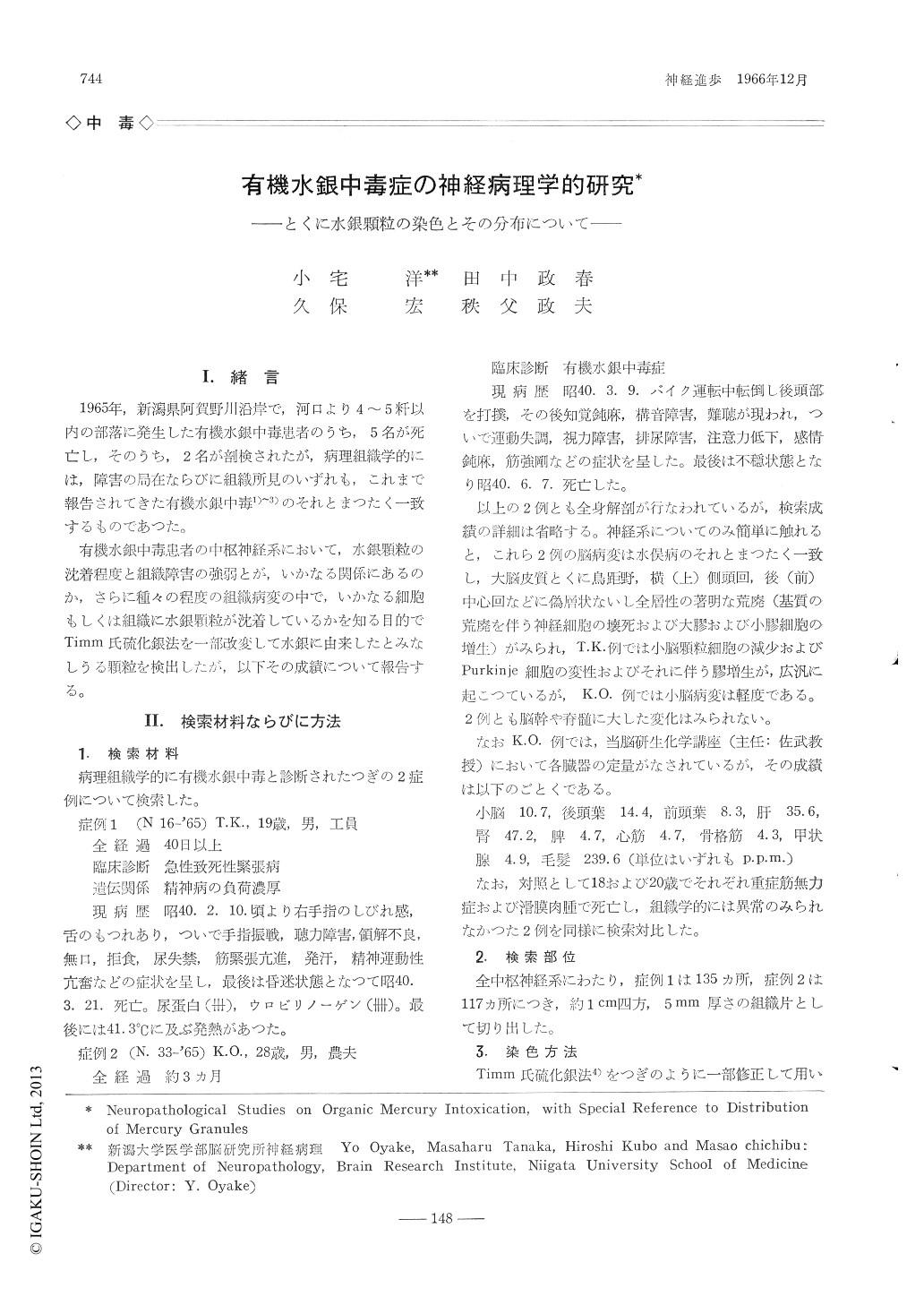
1965年,新潟県阿賀野川沿岸で,河口より4〜5粁以内の部落に発生した有機水銀中毒患者のうち,5名が死亡し,そのうち,2名が剖検されたが,病理組織学的には,障害の局在ならびに組織所見のいずれも,これまで報告されてきた有機水銀中毒1)〜3)のそれとまつたく一致するものであつた。
有機水銀中毒患者の中枢神経系において,水銀顆粒の沈着程度と組織障害の強弱とが,いかなる関係にあるのか,さらに種々の程度の組織病変の中で,いかなる細胞もしくは組織に水銀顆粒が沈着しているかを知る目的でTimm氏硫化銀法を一部改変して水銀に由来したとみなしうる顆粒を検出したが,以下その成績について報告する。
Two cases (Case 1: a 19-year-old male wor-ker, with over 40 day's clinical course. Case 2: a 28-year-old male farmer, with 3 months's clinical course) of organic mercury intoxication, occurred in Niigata district in 1965, were histologically and histochemically examined. Histologic findings of the two were very similar to those of Minamata disease, i.e. cortices of calcarine area, transverse temporal and post-central gyri showed marked devastation (neu-ronal loss, astrocytic and microglial prolifera-tion, and spongiose interstice). Devastation of granule cell layer of cerebellum was prominent in case 1, but slight in case 2.
Over 100 small tissue pieces were taken from various sites of brain and cord and impregnated with Timm's method (slightly modified by us) for heavy metals. Developed granules, which were thought to be mostly mercury granules with some reasons, were found mainly in astro-cytes, microglia and adventitial cells and few, if any, neurons. Distribution of granules was plotted in central nervous system (Photo 2 and 3). Heavy deposition occurred in calcarine cortex, transverse temporal and postcentral gyri as well as cerebellar cortex, corresponding so exactly to areas with heavy pathologic changes. This finding may suggest an importan-ce of histotoxic effect of agens, with regard to pathogenesis of organic mercury intoxication.

Copyright © 1966, Igaku-Shoin Ltd. All rights reserved.


