Japanese
English
- 有料閲覧
- Abstract 文献概要
- 1ページ目 Look Inside
Ⅰ.緒言
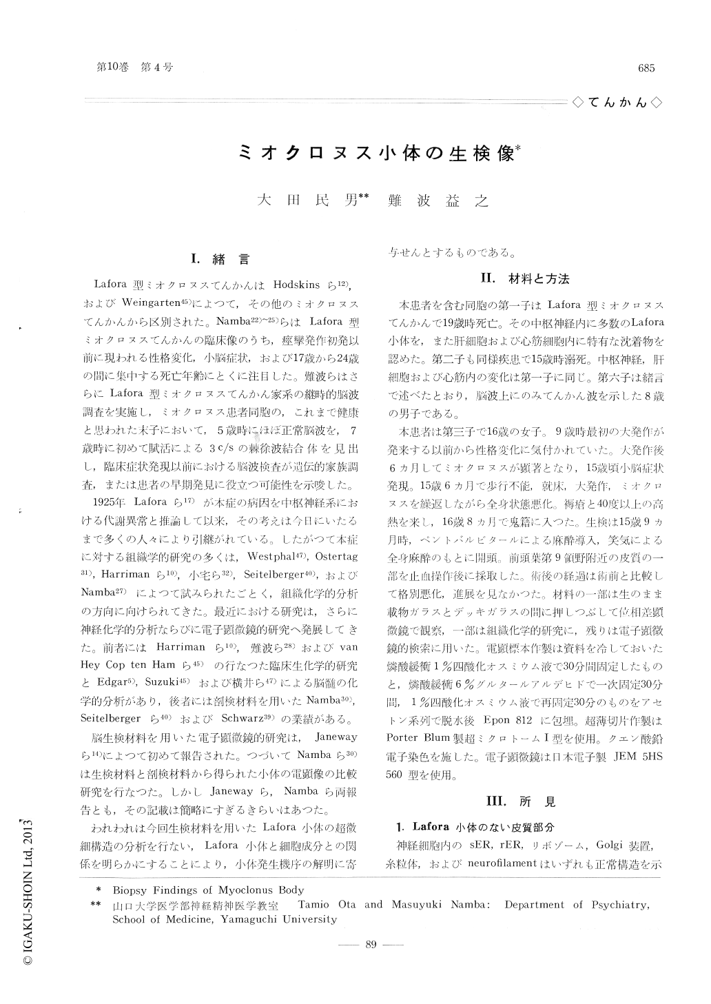
Lafora型ミオクロヌスてんかんはHodskinsら12),およびWeingarten45)によつて,その他のミオクロヌスてんかんから区別された。Namba22)〜25)らはLafora型ミオクロヌスてんかんの臨床像のうち,痙攣発作初発以前に現われる性格変化,小脳症状,および17歳から24歳の間に集中する死亡年齢にとくに注目した。難波らはさらにLafora型ミオクロヌスてんかん家系の継時的脳波調査を実施し,ミオクロヌス患者同胞の,これまで健康と思われた末子において,5歳時にほぼ正常脳波を,7歳時に初めて賦活による3c/sの棘徐波結合体を見出し,臨床症状発現以前における脳波検査が遺伝的家族調査,または患者の早期発見に役立つ可能性を示唆した。
1925年Laforaら17)が本症の病因を中枢神経系における代謝異常と推論して以来,その考えは今日にいたるまで多くの人々により引継がれている。したがつて本症に対する組織学的研究の多くは,Westphal47),Ostertag31),Harrimanら10),小宅ら32),Seitelberger40),およびNamba27)によつて試みられたごとく,組織化学的分析の方向に向けられてきた。
Brain biopsy from 15-year-old girl patient suffering from myoclonus epilepsy (Lafora type) was performed.
Biopsied specimen was studied by the light microscope, the phase contrast microscope and the electron microscope.
Light microscopic examination revealed many Lafora bodies and was completely consistent with the autopsy finding of siblings, already died of myoclonus epilepsy of Lafora type.
Phase contrast microscopic study disclosed the body consisted of two different layers, i. e. the external and the internal one.
Electron micrograph of Lafora body revealed that its basic structure was consisted of lamella which composed of double electron dense lines measured about 50 to 100 A thick and electron lucent interspace of similar thickness. Glycogen-like granules of about 250 to 500 A in diameter were scattered throughout the body. Beside the basic lamellar structure, ribosomes, vacuolated rough surfaced endoplasmic reticulum and Golgi apparatus were also found in the large sized body.
Discussions were performed concerning following items.
(1) cortex without Lafora body.
(2) Lafora body in the neuronal perikarya.
(3) small sized Lafora body.
(4) structures simulate to Lafora body.
The relation of abnormal vesicle and the process of Lafora body formation was especially discussed.

Copyright © 1966, Igaku-Shoin Ltd. All rights reserved.


