Japanese
English
- 有料閲覧
- Abstract 文献概要
- 1ページ目 Look Inside
はじめに
コンピューター断層撮影,Computed Tomo-graphy(以下CTと略す)の泌尿器科的応用は,全身用装置の普及とともにもはや日常検査の一つとなりつつある1,2)。
われわれは先に本誌に第2世代のCTスキャナーといわれる米国Ohio-nuclear社のDelta-scan50装置による画像を中心に報告した3,4)。なお,各科領域における報告も数多くなされている5,6)。今回は,その後開発された同社のいわゆる第4世代装置,Delta-scan 2020,fan beam X-ray rota-tionary方式を使用する機会を得たので報告する。症例は1979年2月から6月末まで12例経験した。
Twelve cases of urological patients were observed by computed tomography (Ohionuclear's Delta-scan 2020).
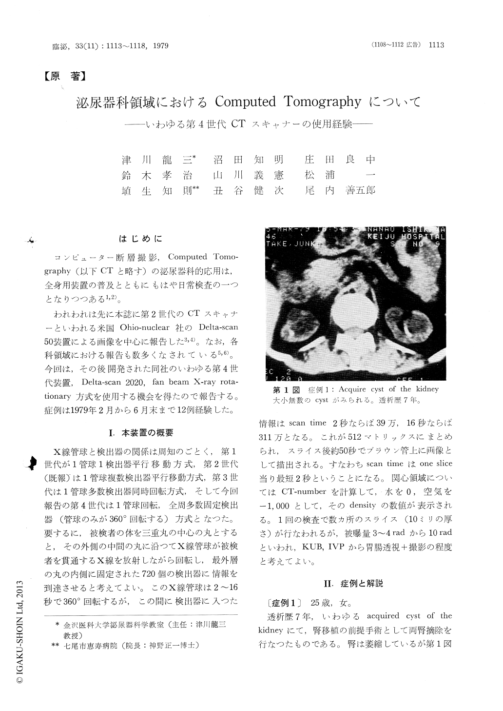
5 cases of them were as follows : 1) A 25-year-old female of chronic renal failure, in which both kidneys showed multiple cystic structure, what is called "acquired cyst of the kidney".
2) A 28-year-old male of chronic renal failure, in which round calcificated shadow was revealed at the left kidney. It was very difficult to demonstrate the round shadow on KUB. Renal carcinoma was suspected by selective renal arteriography, and the nephrectomy specimen revealed renal cell carcinoma.

Copyright © 1979, Igaku-Shoin Ltd. All rights reserved.


