Japanese
English
- 有料閲覧
- Abstract 文献概要
- 1ページ目 Look Inside
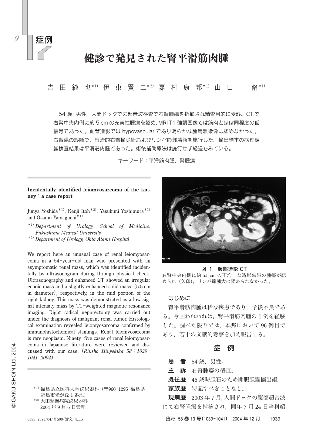
54歳,男性。人間ドックでの超音波検査で右腎腫瘍を指摘され精査目的に受診。CTで右腎中央内側に約5cmの充実性腫瘍を認め,MRI T1強調画像では筋肉とほぼ同程度の低信号であった。血管造影ではhypovascularであり明らかな腫瘍濃染像は認めなかった。右腎癌の診断で,根治的右腎摘除術およびリンパ節郭清術を施行した。摘出標本の病理組織検査結果は平滑筋肉腫であった。術後補助療法は施行せず経過をみている。
We report here an unusual case of renal leiomyosarcoma in a 54-year-old man who presented with an asymptomatic renal mass,which was identified incidentally by ultrasonogram during through physical check. Ultrasonography and enhanced CT showed an irregular echoic mass and a slightly enhanced solid mass(5.5cm in diameter),respectively,in the mid portion of the right kidney. This mass was demonstrated as a low signal intensity mass by T1-weighted magnetic resonance imaging. Right radical nephrectomy was carried out under the diagnosis of malignant renal tumor. Histological examination revealed leiomyosarcoma confirmed by immunohistochemical stainings. Renal leiomyosarcoma is rare neoplasm. Ninety-five cases of renal leiomyosarcoma in Japanese literature were reviewed and discussed with our case.

Copyright © 2004, Igaku-Shoin Ltd. All rights reserved.


