Japanese
English
- 有料閲覧
- Abstract 文献概要
- 1ページ目 Look Inside
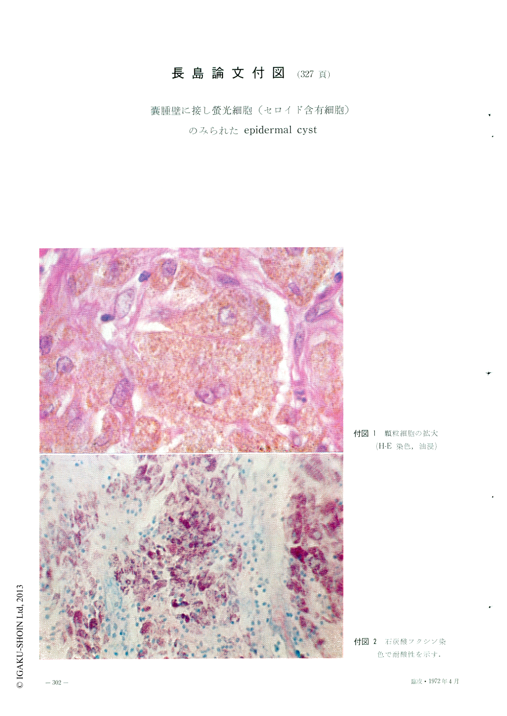
1970年,本間1)は嚢腫壁に接して多数の顆粒細胞のみとめられたepidermal cystの1例を記載し,この顆粒細胞がHamperl2)のいうFluorocyten(螢光細胞)に一致するものであり,しかもこの細胞に含まれている顆粒が,Lillieら3,4)のいうセロイド(ceroid)色素に相当することを組織化学的に証明した.
従来,病理組織学的にepidermal cystの周囲に異物反応がしばしばみとめられることはよく知られており,また日常よく経験される所でもある.しかしながら,従来の成書に該部におけるセロイド色素の出現を指摘したものはない.
A case of this disease in a 34-year-old man was reported. It was a hen-egg-sized tumor on the right buttock with a 7 years' history.
Histologic specimen revcaled many granular cells in the connective tissue surrounding the cystic wall. The granule emitted a yellow-green fluorescence under the fluorescent microscope. It was positive (acid fast) by the carbol-fuchsin stain, strong positive by PAS stain, and positive by Sudan III and Sudanblack B in paraffin section. By these results it was identified as ceroid pigment.
Three in 88 specimen of the epidermal cyst including the author's case for recent 11 years had the ceroid containing cells.

Copyright © 1972, Igaku-Shoin Ltd. All rights reserved.


