Japanese
English
- 有料閲覧
- Abstract 文献概要
- 1ページ目 Look Inside
1962年,FalckおよびHillarpら1,2)は組織を凍結乾燥し,これをformaldehyde gasにて処理することにより,全ての組織におけるCatecholamines(CAと略記)および5-Hydroxytryptamine(Serotonin)(5-HTと略記)の局在を細胞ないし細胞下レベルで形態学的に追求可能とした。さらに,Corrodi & Jonsson3)によつて,その化学反応が明らかにされ,またThieme4)は螢光顕微分光法により,該螢光がauthentic NAと完全に一致することを証明するに至り,本法の特異性に関する信頼度が一躍増大した。
かかる螢光法を用いての皮膚モノアミン作動神経の研究はFalckら5),Moller6)および森嶋7)により,またメラノサイト,母斑細胞ならびに悪性黒色腫細胞の観察はFalckら8,9),橋本ら10,11)および森嶋,遠藤21)によりなされている。
Histochemical studies by fluorescent technique on the typical malignant melanoma, which has developed on the inner surface of the left arm of a 32-year-old woman for 2 years and was a darkbrawn, 1.5×1.8cm in size, were performed.
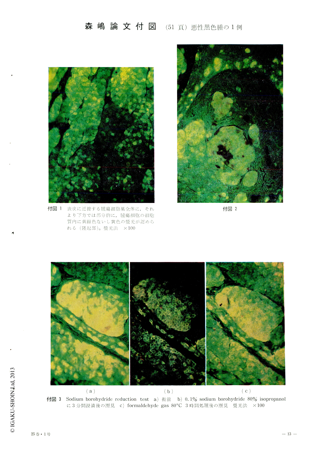
A green, yellowgreen or yellow fluorescence was observed in the cytoplasm of the tumor cells treated with formaldehyde. It was concluded that the fluorescence should be due to the first order of cathecolamine or its precursor of-DOPA. The reasons which gave this conclusion arc as follows:
1) The specimen was treated with formaldehyde gas with 60% humidity, at 80℃, for 1 hour.
2) The fluorescence was detected by the ultraviolet of the light source of the fluorescent microscope.
3) It was quenched by the immersion into the 40% isopropanol solution.
The differentiation among noradrenalin, DOPA amine and DOPA was difficult, because these substances had the same emission and excitation spectrum for the fluorescence.
Because the results of the thionyl chloride test, the biochemical analysis of the melanoma, and lack DOPA decarboxylase in the mouse melanoma, the author suspected the specific fluorescence of the melanoma cells might be due to mainly DOPA.

Copyright © 1971, Igaku-Shoin Ltd. All rights reserved.


