Japanese
English
- 有料閲覧
- Abstract 文献概要
- 1ページ目 Look Inside
1915年Fabry1)がそれまでAngiokeratomaとして片付けられていた疾患の内,母斑性でしかも躯幹または四肢の片側に存在するものに対してAngiokeratoma circumscriptum naeviformeなる名称のもとに1独立疾患として報告した。その後本症に一致すると思われる疾患に対して,keratotic hernangiorna2),hemangioma verrucosum3),naevus angiokeratoticus10),その他種々の名で発表されてきた。
本症の報告数は外国で40例内外4),本邦で17例内外6〜18)(表参照)に過ぎず,非常にまれな疾患と思われるが,それは単なる血管腫として見のがされている可能性もある。
Four case of this disease were reported.
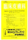
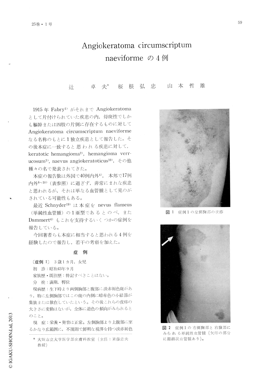
Case 1 : A 3-year-and-10-month-old girl showed a large brownred macula with many clarkred nodules on the left sidechest and epigastrial region. She also had portwinc stains on the right sidechest and the right epigastrial region, and cavernous angiomas on the right axilla and the right sidechest.
Case 2 : A 3-year-and-7-month-old boy had small verrucous, miliar to peasized, yellow-white or dark-red nodules on the extensor and inner surfaces of the right thigh. There was a cavernous angioma over the large area around the nodules.
Case 3 : A 10-year-old gial had a dark-red or whitish yellow, verrucous, egg-sized, elevated plaques on the extensor aspect of the left thigh.
Case 4 : A 21-year-old woman had a dark-red, thumbtip-sized, uneven, elevated macula on the flexor aspect of the right thight.
Histologic syccimens of these cases showed almost the same pictures which were typical of this disease. The alkaline phosphatase reaction on the dilated vascular wall was negative. Electron microscopically the abnormally dilated vessels were lined with one layer of the endothelial cell, the subcellular organelles of which had no abnormality.
This disease seemed to be caused by some external stimulation on the hemangioma simplex.
Copyright © 1971, Igaku-Shoin Ltd. All rights reserved.