Japanese
English
- 有料閲覧
- Abstract 文献概要
- 1ページ目 Look Inside
I.はじめに
我々は今回,約16年前より右踵部に出現し最近徐々に拡大して来た色素斑の1例を経験し,臨床的ならびに組織学的にMelanosis circumscripta precancerosaより発生した悪性黒色腫と診断したのでここに報告する。
A 63-year-old man had pigmented macule on the right heel 16 years ago, which increased its size and had erosion.
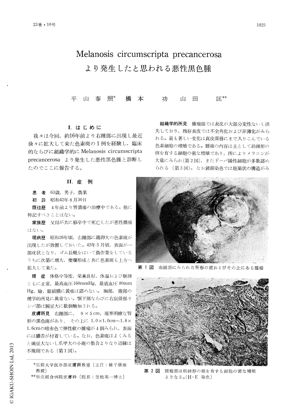
On examination there was a black, kidney-shaped, well margined macule, 5×9 cm in size, with 4, dark-red, elastic hard tumors on it. The macule with irregular border was composed of smaller macules upto nale in size.
In histologic specimen from the tumor epidermis degenerated or disappeared almost entirely, and survived epidermis was thin and parakeratotic. The tumor was composed of cells with spindle shaped nucleus, containing partly many melanin granules, and showing DOPA positive reaction in the most of them.
Specimen from the macule around the tumor showed hyperkeratosis, casting FT of melanin into the horny layer, many melanocytes in the basal cell layer. Papillary layer of the dermis was edematous and showed lymphocytic infiltration and marked junctional activity. Lymphopl-asmacytic infiltrations around the vessels and sweat glands were proved in the middermis. There was no evidence of metastasis in the regional lymphatic systems, including inguinal lymphnodes.
Laboratory tests revealed nothing particular except slight anemia, increased sedimentation rate, and positive STS. No melanuria was proved.
After soft x-ray radiation and anticancer drug therapy, the lesion including large area of normal skin was extripated and grafted.
Postoperative administration of COPP and soft x-ray radiation were performed. There was no evidence of recurrence 9 months after operation.
Copyright © 1969, Igaku-Shoin Ltd. All rights reserved.