Japanese
English
- 有料閲覧
- Abstract 文献概要
- 1ページ目 Look Inside
はじめに
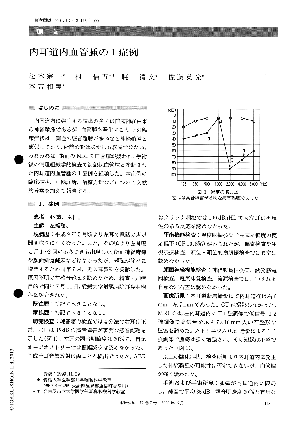
内耳道内に発生する腫瘍の多くは前庭神経由来の神経鞘腫であるが,血管腫も発生する1)。その臨床症状は一側性の感音難聴が多いなど神経鞘腫と類似しており,術前診断は必ずしも容易ではない。われわれは,術前のMRIで血管腫が疑われ,手術後の病理組織学的検査で海綿状血管腫と診断された内耳道内血管腫の1症例を経験した。本症例の臨床症状,画像診断,治療方針などについて文献的考察を加えて報告する。
A 45-year-old female had suffered from a pro-gressing hearing loss of the left ear for 2 months. Pure tone audiogram showed a sensorineural hear-ing loss of 35 dB. A small tumor, 7×10mm in diameter was revealed in the internal auditory meatus by MRI. The tumor represented a low inten-sity on T 1-weight image, a high intensity on T 2-weight image and positive Gd enhancement of T 1-weight image. The tumor was totally removed by the middle cranial fossa approach with preservation of the facial nerve function, however her hearing deteriorated postoperatively. Histologically the tumor was cavernous hemangioma.

Copyright © 2000, Igaku-Shoin Ltd. All rights reserved.


