Japanese
English
- 有料閲覧
- Abstract 文献概要
- 1ページ目 Look Inside
はじめに
生前の聴覚機能と内耳病態の比較病理組織学的研究は,難治な内耳疾患の解明とその臨床的対応に結びつくためにかねてからかなりの報告がある1)。老人性難聴に関しては,蝸牛の加齢による病理学的変化が単一ではなく,症例によって蝸牛における種々の部位に特徴的な病的変化を生じるところから,1) sensory presbycusis,2) neural presbycusis,3) strial presbycusis,4) coch-lear conductive presbycusisの4つの代表的な病型に分類され1),聴覚所見との関係が論じられている。
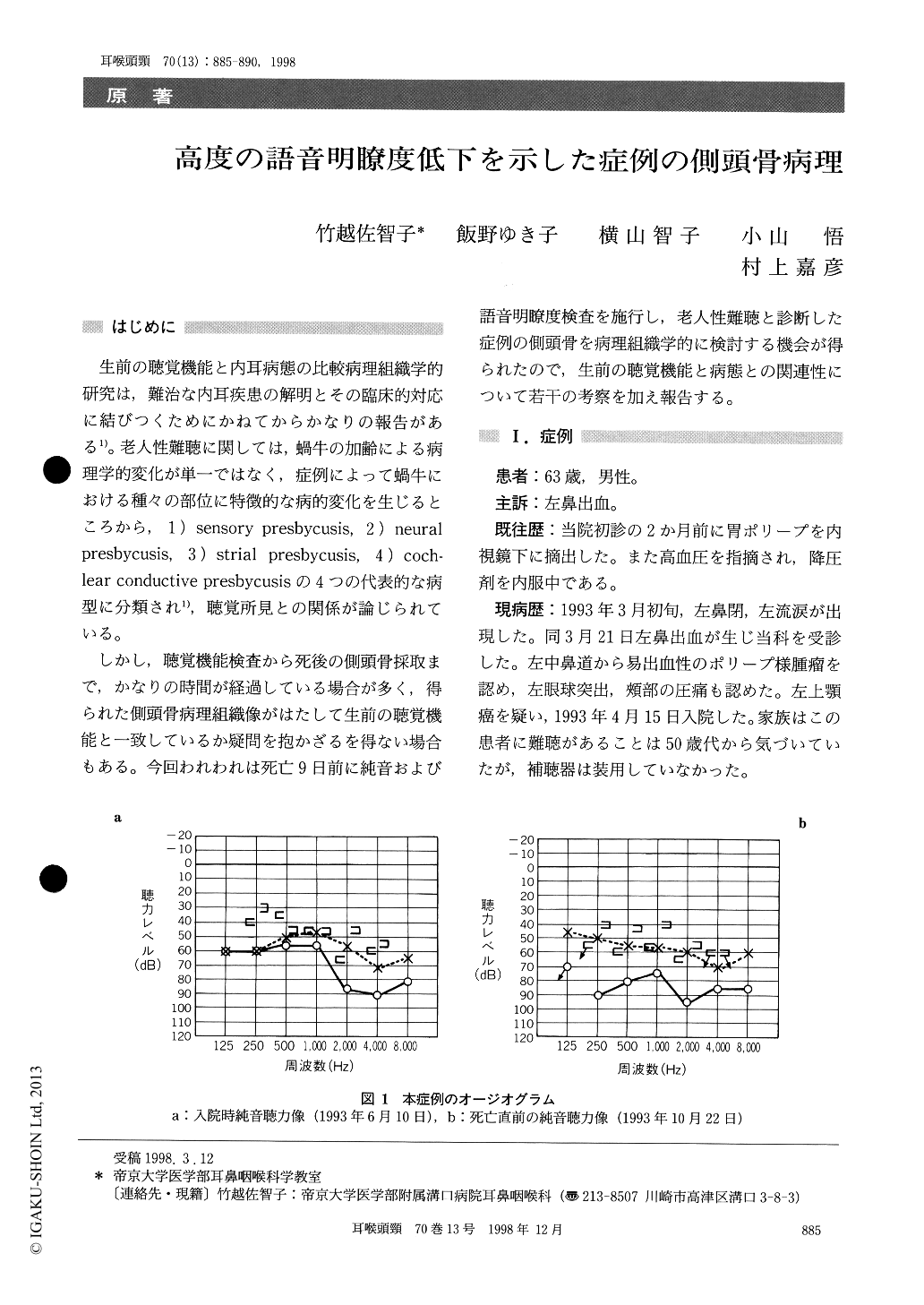
しかし,聴覚機能検査から死後の側頭骨採取まで,かなりの時間が経過している場合が多く,得られた側頭骨病理組織像がはたして生前の聴覚機能と一致しているか疑問を抱かざるを得ない場合もある。今回われわれは死亡9日前に純音および語音明瞭度検査を施行し,老人性難聴と診断した症例の側頭骨を病理組織学的に検討する機会が得られたので,生前の聴覚機能と病態との関連性について若干の考察を加え報告する。
We report the temporal bone histopathology in a 63-year-old male with a moderate sensorineural hearing loss and poor speech discrimination. His hearing acuity was evaluated by pure tone andspeech audiometry 10 days before his death. A histological study on the temporal bones revealed marked loss of the spiral ganglion cells and den-drites in all cochlear turns. The neuronal loss was mostly limited to the afferent nerve fibers, while the efferent nerve fibers were spared. The outer and inner hair cells as well as the stria vascularis appeared almost normal. From these temporal bone findings, the sensorineural hearing loss in this patient was diagnosed as neural presbycusis of Schuknecht.

Copyright © 1998, Igaku-Shoin Ltd. All rights reserved.


