Japanese
English
- 有料閲覧
- Abstract 文献概要
- 1ページ目 Look Inside
はじめに
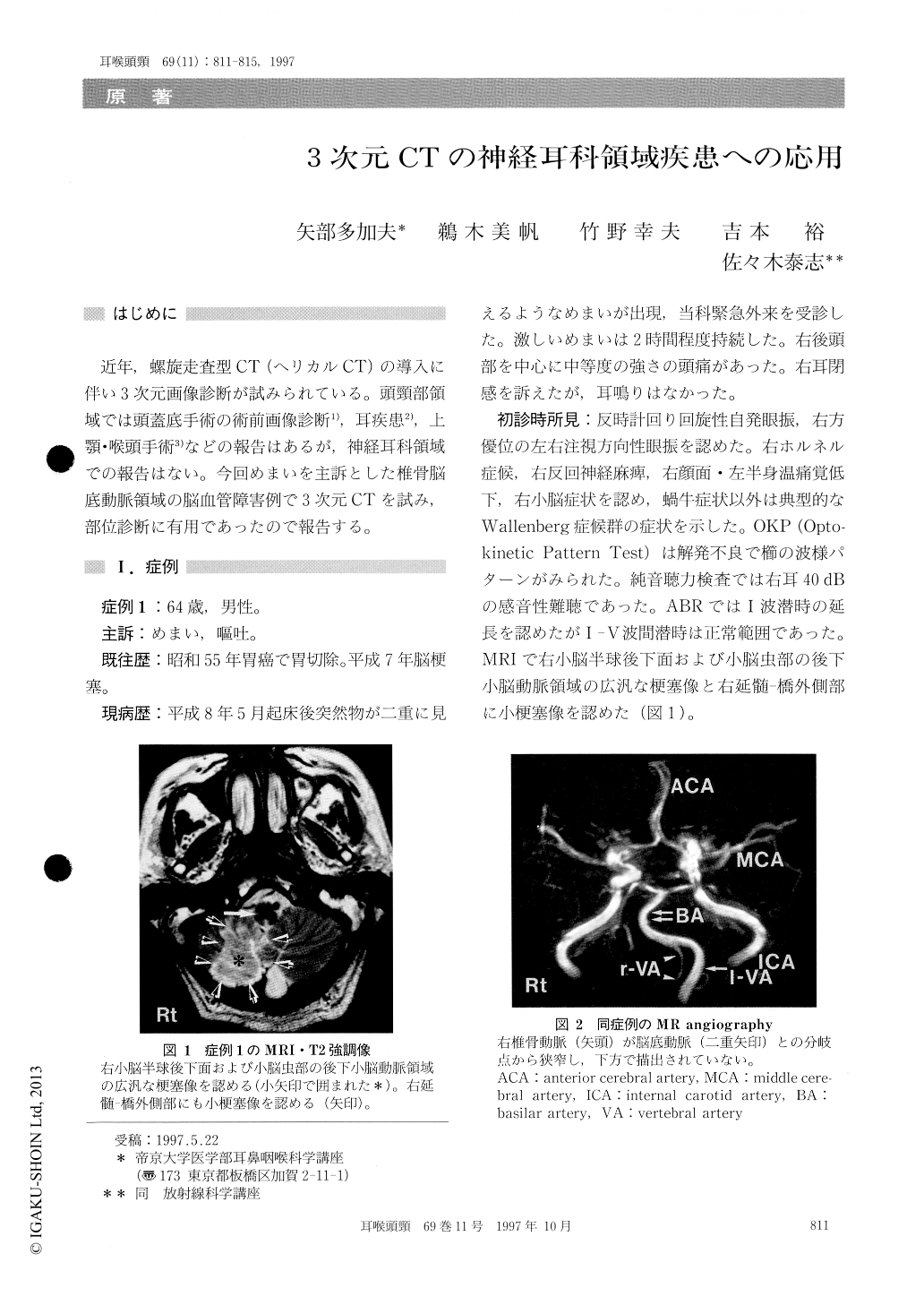
近年,螺旋走査型CT (ヘリカルCT)の導入に伴い3次元画像診断が試みられている。頭頸部領域では頭蓋底手術の術前画像診断1),耳疾患2),上顎・喉頭手術3)などの報告はあるが,神経耳科領域での報告はない。今回めまいを主訴とした椎骨脳底動脈領域の脳血管障害例で3次元CTを試み,部位診断に有用であったので報告する。
In two patients of cerebellar vascular infarction who had a complaint of vertigo, the clinical applica-tion of 3D CT for neurotological disorders was discussed. Case 1 : a 64-year-old male. MRI revealed extensive infarct lesions of the posterior cerebellar artery (PICA) and 3D-CT demonstrated a occlusive lesion of the right vertebral artery. Case 2 : a 55-year-old male. MRI revealed localized in-farct lesions of the cerebellar vermis and from 3D-CT the occlusive lesions of the medial branch of the right PICA were suspected. 3D-CT was a usefull diagnostic tool to detect the site of vascular lesions.

Copyright © 1997, Igaku-Shoin Ltd. All rights reserved.


