Japanese
English
- 有料閲覧
- Abstract 文献概要
- 1ページ目 Look Inside
はじめに
錐体骨から発生する腫瘍性病変はある程度の大きさになって内耳あるいは内耳道に進展すると,はじめて顔面神経麻痺,感音難聴,めまいといった症状を起こしてくるが,単純耳X線撮影,内耳道断層撮影では骨破壊像をとらえることがなかなか難しい。同様の症状を示す他疾患との鑑別が困難て,発見も遅れがちとなり,CT, MRIを行ってようやく診断がつくことが多い。そのため発症から診断,手術までの経過が長く,神経変性も高度となって,予後不良となりやすい。さらに部位的に手術操作が困難で全摘術が難しいことも多い。
今回われわれは,難聴と顔面神経麻痺を伴った錐体骨原発と思われる類上皮腫と両側の嚢胞性腫瘍を経験したので,文献的考察を加え報告する。
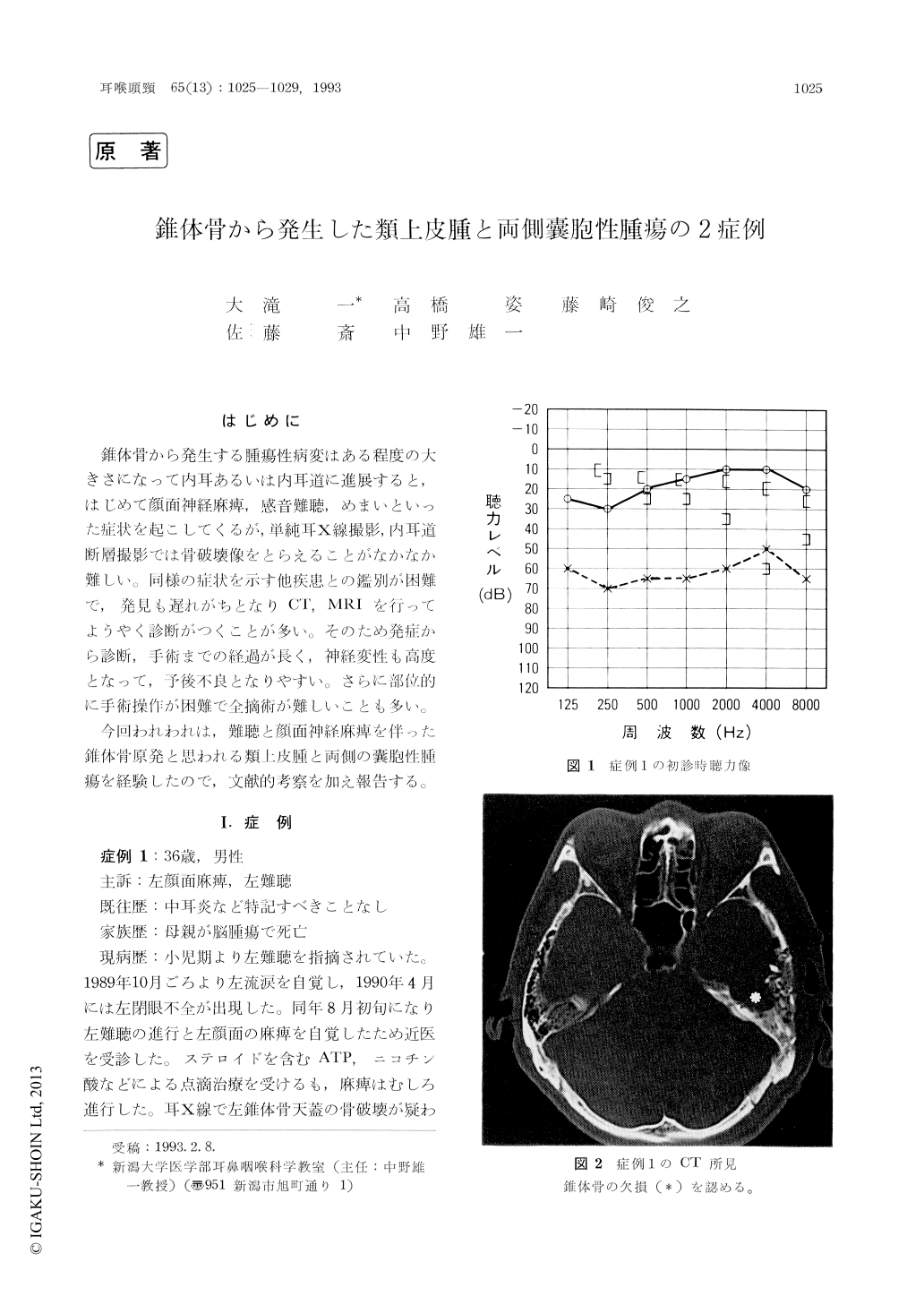
Case 1: A 35-year-old man presented with a long history of slowly progressed left hearing loss and recent facial paralysis. A large epidermoid tumor associated with a left petrous bone defect was detected by CT and MRI.
Case 2: An 18-year-old woman presented with a fluctuating bilateral hearing loss and right facial paralysis. CT showed bilateral petrous bone defects and MRI revealed a cystic tumor in each side.
Epidermoid tumor in case 1 and the right cystic tumor in case 2 were removed by trans-middle cranial fossa approach and trans-posterior cranial fossa approach, respectively.
The usefulness of CT and MRI images in eva-luating petrous bone lesions was confirmed.

Copyright © 1993, Igaku-Shoin Ltd. All rights reserved.


