Japanese
English
- 有料閲覧
- Abstract 文献概要
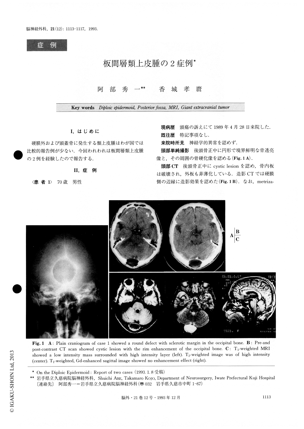
- 1ページ目 Look Inside
I.はじめに
硬膜外および頭蓋骨に発生する類上皮腫はわが国では比較的報告例が少ない.今回われわれは板間層類上皮腫の2例を経験したので報告する.
Primary epidermoid tumors comprise about 1% of all central nervous system neoplasms, although the diploic epidermoid tumor is comparatively rare. Two cases of di-ploic epidermoid tumor are reported in this paper.
Case 1 : A 70-year-old man presented with a headache. A plain craniogram showed an osteolytic lesion of the occipital bone with a well defined sclerotic margin. A contrast enhanced CT confirmed a cystic lesion with rim enhancement. On MRI, the tumor appeared hypoin-tense surrounded with irregular hyperintensity on the T1WI and hyperintensity on the T2WI. Gd enhancement on the MRI showed no enhancement effect. The tumor was totally removed and cranioplasty was performed. No tumor invasion of the dura mater was noticed.
Case 2 : A 90-year-old woman presented with a giant tumor of the left parietal region. She noticed a painless swelling at the age of 20, and the tumor slowly grew over a period of 70 years. Plain craniogram showed a bony de-fect with a sclerotic margin. CT scan confirmed an ex-tracranial giant tumor with destruction of the outer table under the tumor, and also falx meningioma. Aspiration and irrigation inside the cystic tumor were performed under local anesthesia.
Previous authors have also said that the plain cranio-gram is characteristic and diagnostic in the case of dip-loic epidermoid. Typical round or polylobular bony de-fect with well defined sclerotic margins was visualized.
Copyright © 1993, Igaku-Shoin Ltd. All rights reserved.