Japanese
English
- 有料閲覧
- Abstract 文献概要
- 1ページ目 Look Inside
I.はじめに
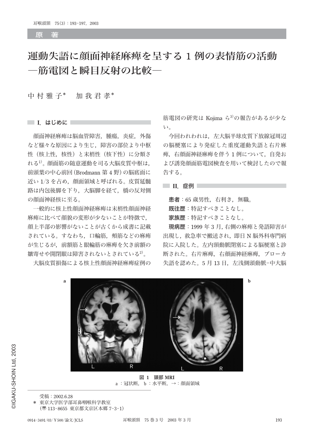
顔面神経麻痺は脳血管障害,腫瘍,炎症,外傷など様々な原因により生じ,障害の部位より中枢性(核上性,核性)と末梢性(核下性)に分類される1)。顔面筋の随意運動を司る大脳皮質中枢は,前頭葉の中心前回(Brodmann第4野)の脳底面に近い1/3を占め,顔面領域と呼ばれる。皮質延髄路は内包後脚を下り,大脳脚を経て,橋の反対側の顔面神経核に至る。
一般的に核上性顔面神経麻痺は末梢性顔面神経麻痺に比べて顔貌の変形が少ないことが特徴で,顔上半部の影響がないことが古くから成書に記載されている。すなわち,口輪筋,頬筋などの麻痺が生じるが,前額筋と眼輪筋の麻痺を欠き前額の皺寄せや開閉眼は障害されないとされている2)。
大脳皮質損傷による核上性顔面神経麻痺症例の筋電図の研究はKojimaら3)の報告があるが少ない。
今回われわれは,左大脳半球皮質下放線冠周辺の脳梗塞により発症した重度運動失語と右片麻痺,右顔面神経麻痺を伴う1例について,自発および誘発顔面筋電図検査を用いて検討したので報告する。
A patient presented with supranuclear facial palsy after cerebral infarction of the left frontal lobe. The facial palsy was moderate degree in the right frontal belly,orbicularis oculi and orbicularis oris muscles. However,the difference was not recognized on spontaneous electromyogram and evoked electromyogram. However it was found that there was a low amplitude of R2 in the affected side by stimulation and healthy side of the right and left by stimulation in the affected side in blink reflex. This low R2 was thought to be due to the damage of the superior nucleus of the reflex pathway of R2.

Copyright © 2003, Igaku-Shoin Ltd. All rights reserved.


