Japanese
English
- 有料閲覧
- Abstract 文献概要
- 1ページ目 Look Inside
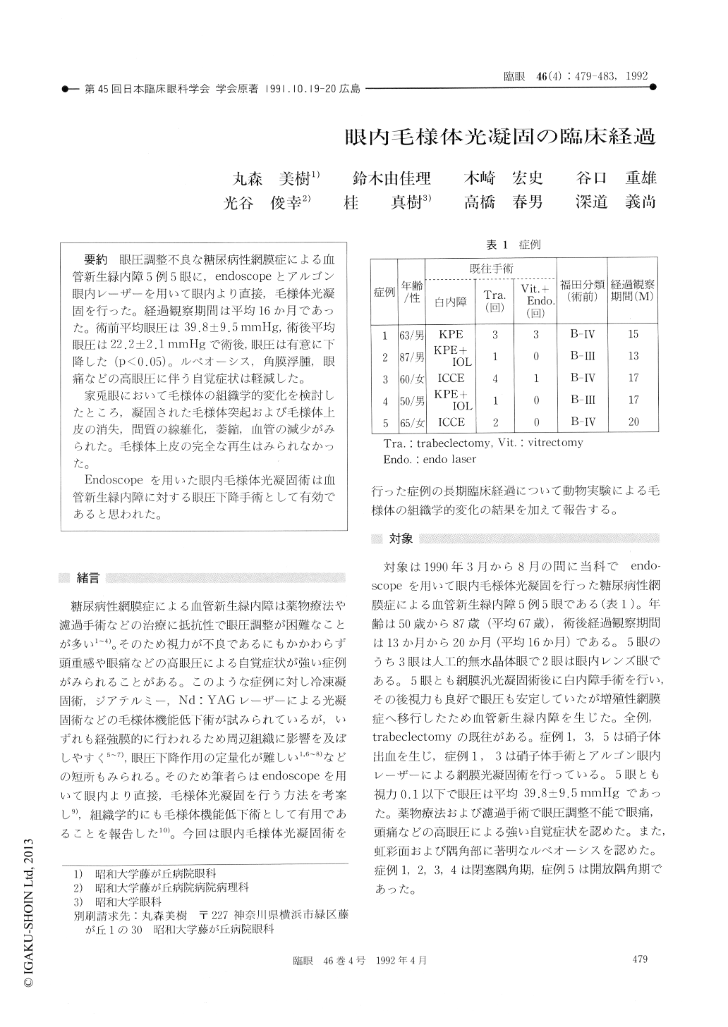
眼圧調整不良な糖尿病性網膜症による血管新生緑内障5例5眼に,endoscopeとアルゴン眼内レーザーを用いて眼内より直接,毛様体光凝固を行った。経過観察期間は平均16か月であった。術前平均眼圧は39.8±9.5mmHg,術後平均眼圧は22.2±2.1mmHgで術後,眼圧は有意に下降した(p<0.05)。ルベオーシス,角膜浮腫,眼痛などの高眼圧に伴う自覚症状は軽減した。
家兎眼において毛様体の組織学的変化を検討したところ,凝固された毛様体突起および毛様体上皮の消失,間質の線維化,萎縮,血管の減少がみられた。毛様体上皮の完全な再生はみられなかった。
Endoscopeを用いた眼内毛様体光凝固術は血管新生緑内障に対する眼圧下降手術として有効であると思われた。
We performed argon laser photocoagulation of the ciliary body in 5 eyes of 5 diabetic patients with neovascular glaucoma and poorly controlled intraocular pressure (IOP). The ciliary proceses were coagulated under direct endoscopic observa-tion. The area of coagulation ranged from 120 to 240 degrees, average 156 degrees. The follow-up ranged 13 to 20 months, average 16 months. The IOP averaged 39.8 mmHg before and 22.2 mmHg after treatment. In all the 5 eyes, neovasculariza-tion on the iris and chamber angle regressed or disappeared after treatment. Slight vitreous hemor-rhage developed in 2 eyes but resolved spontaneous-ly. There was no instance of postoperative ocular hypotension or phthisis bulbi. Studies in 20 pigment-ed rabbit eyes after similar treatment showed dis-appearance of ciliary folds, destruction of epithelial cells and atrophy of ciliary stroma in photo-coagulated areas. The findings show the efficacy of endoscopic cyclophotocoagulation in diabetic eyes with poorly controlled neovascular glaucoma.

Copyright © 1992, Igaku-Shoin Ltd. All rights reserved.


