Japanese
English
- 有料閲覧
- Abstract 文献概要
- 1ページ目 Look Inside
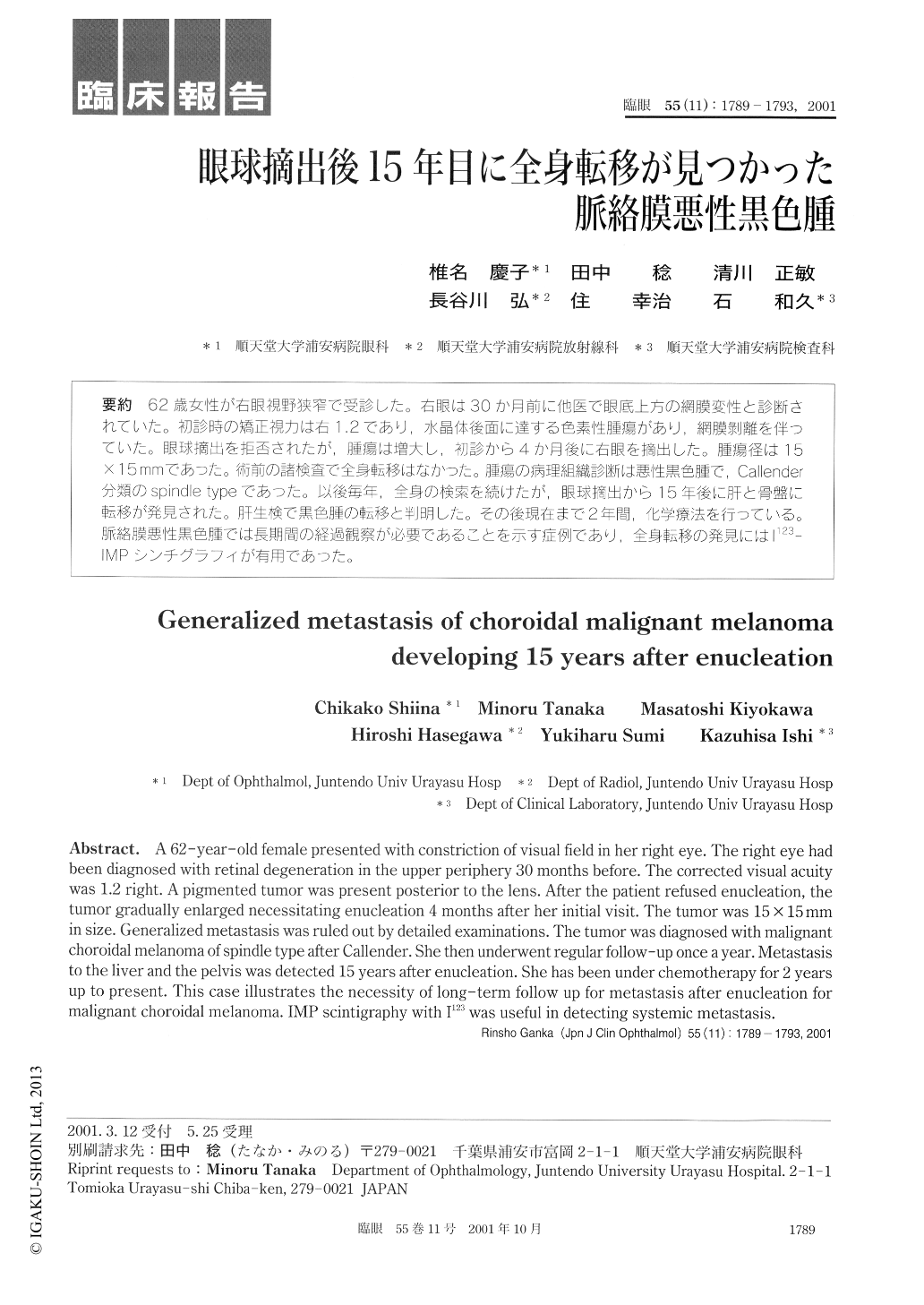
62歳女性が右眼視野狭窄で受診した。右眼は30か月前に他医で眼底上方の網膜変性と診断されていた。初診時の矯正視力は右1.2であり,水晶体後面に達する色素性腫瘍があり,網膜剥離を伴っていた。眼球摘出を拒否されたが,腫瘍は増大し,初診から4か月後に右眼を摘出した。腫瘍径は15×15mmであった。術前の諸検査で全身転移はなかった。腫瘍の病理組織診断は悪性黒色腫で,Callender分類のspindle typeであった。以後毎年,全身の検索を続けたが,眼球摘出から15年後に肝と骨盤に転移が発見された。肝生検で黒色腫の転移と判日月した。その後現在まで2年間,化学療法を行っている。脈絡膜悪性黒色腫では長期間の経過観察が必要であることを示す症例であり,全身転移の発見にはI123—IMPシンチグラフィが有用であった。
A 62-year-old female presented with constriction of visual field in her right eye. The right eye had been diagnosed with retinal degeneration in the upper periphery 30 months before. The corrected visual acuity was 1.2 right. A pigmented tumor was present posterior to the lens. After the patient refused enucleation, the tumor gradually enlarged necessitating enucleation 4 months after her initial visit. The tumor was 15×15mm in size. Generalized metastasis was ruled out by detailed examinations. The tumor was diagnosed with malignant choroidal melanoma of spindle type after Callender. She then underwent regular follow-up once a year. Metastasis to the liver and the pelvis was detected 15 years after enucleation. She has been under chemotherapy for 2 years up to present. This case illustrates the necessity of long-term follow up for metastasis after enucleation for malignant choroidal melanoma. IMP scintigraphy with I123 was useful in detecting systemic metastasis.

Copyright © 2001, Igaku-Shoin Ltd. All rights reserved.


