Japanese
English
- 有料閲覧
- Abstract 文献概要
- 1ページ目 Look Inside
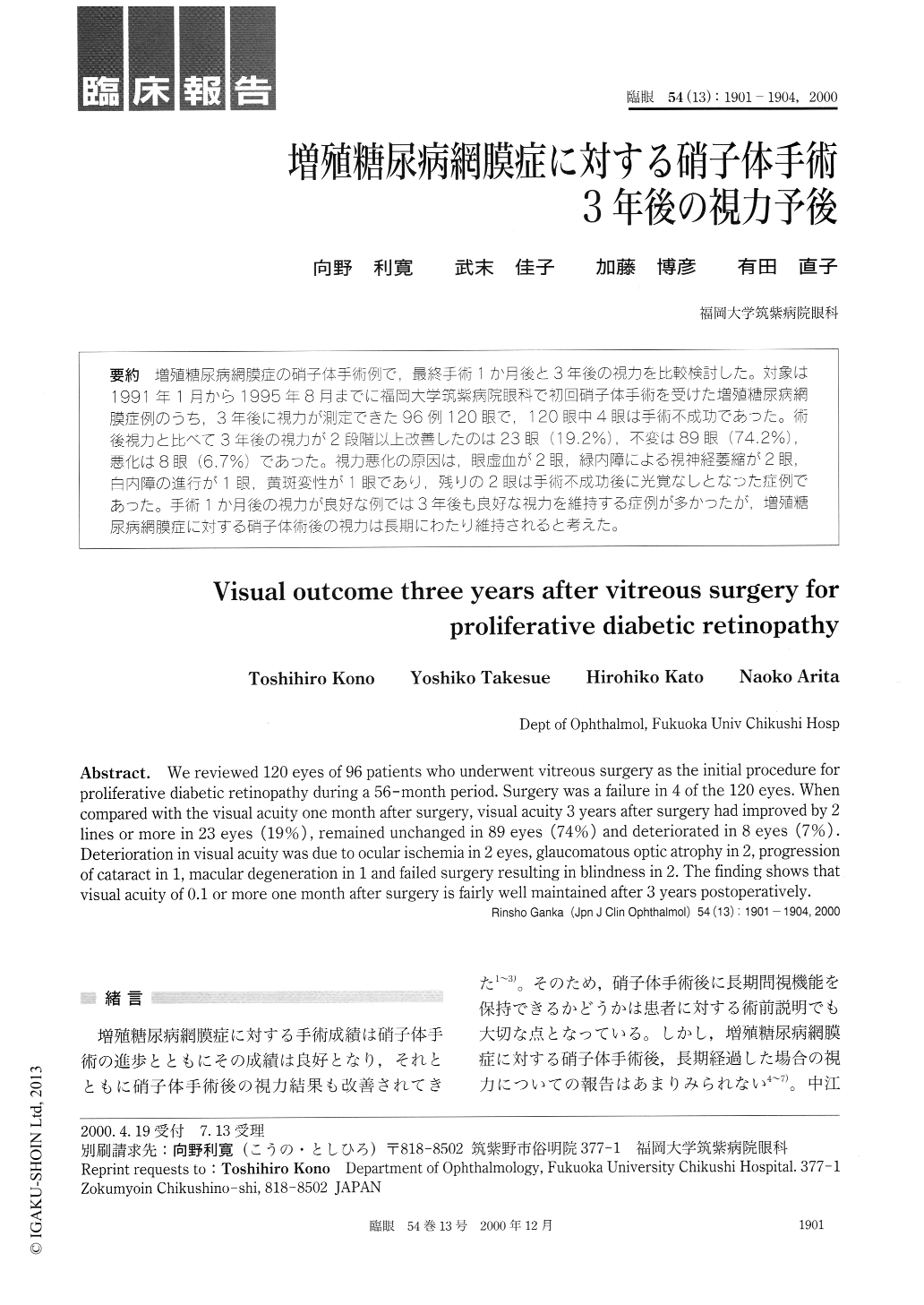
増殖糖尿病網膜症の硝子体手術例で,最終手術1か月後と3年後の視力を比較検討した。対象は1991年1月から1995年8月までに福岡大学筑紫病院眼科で初回硝子体手術を受けた増殖糖尿病網膜症例のうち,3年後に視力が測定できた96例120眼で,120眼中4眼は手術不成功であった。術後視力と比べて3年後の視力が2段階以上改善したのは23眼(9.2%),不変は89眼(74.2%),悪化は8眼(6.7%)であった。視力悪化の原因は,眼虚血が2眼,緑内障による視神経萎縮が2眼,白内障の進行が1眼,黄斑変性が1眼であり,残りの2眼は手術不成功後に光覚なしとなった症例であった。手術1か月後の視力が良好な例では3年後も良好な視力を維持する症例が多かったが,増殖糖尿病網膜症に対する硝子体術後の視力は長期にわたり維持されると考えた。
We reviewed 120 eyes of 96 patients who underwent vitreous surgery as the initial procedure for proliferative diabetic retinopathy during a 56-month period. Surgery was a failure in 4 of the 120 eyes. When compared with the visual acuity one month after surgery, visual acuity 3 years after surgery had improved by 2 lines or more in 23 eyes (19%) , remained unchanged in 89 eyes (74%) and deteriorated in 8 eyes (7%) . Deterioration in visual acuity was due to ocular ischemia in 2 eyes, glaucomatous optic atrophy in 2, progression of cataract in 1, macular degeneration in 1 and failed surgery resulting in blindness in 2. The finding shows that visual acuity of 0.1 or more one month after surgery is fairly well maintained after 3 years postoperatively.

Copyright © 2000, Igaku-Shoin Ltd. All rights reserved.


