Japanese
English
- 有料閲覧
- Abstract 文献概要
- 1ページ目 Look Inside
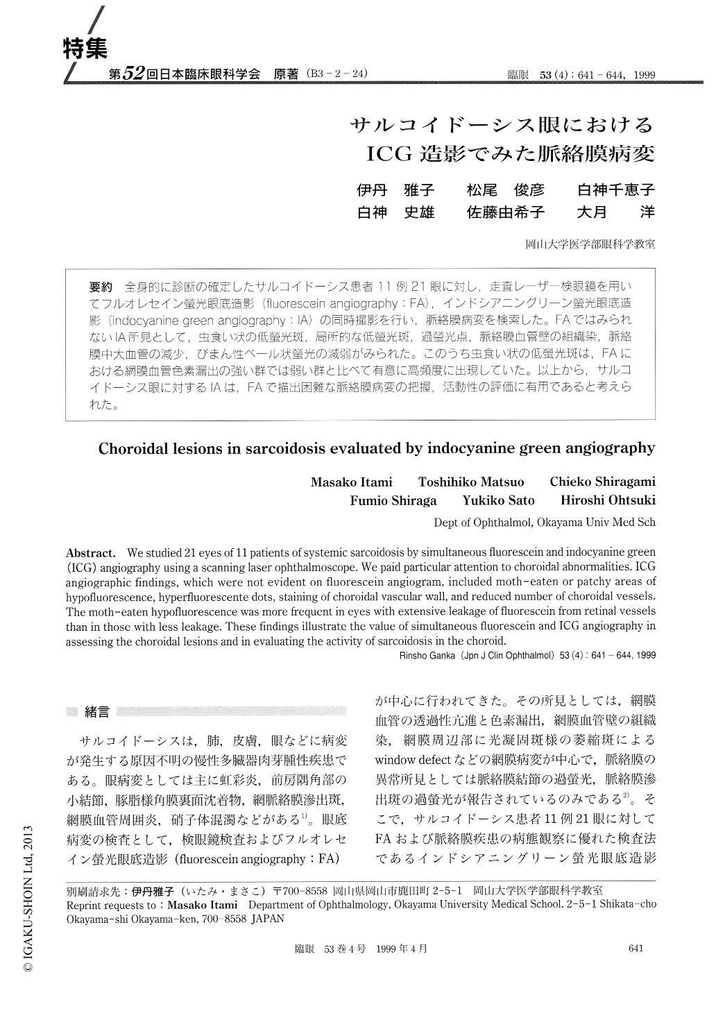
(B3-2-24) 全身的に診断の確定したサルコイドーシス患者11例21眼に対し,走査レーザー検眼鏡を用いてフルオレセイン螢光眼底造影(fluorescein angiography:FA),インドシアニングリーン螢光眼底造影(indocyanhe green angiQgraphy:IA)の同時撮影を行い,脈絡膜病変を検索した。FAではみられないIA所見として,虫食い状の低螢光斑,局所的な低螢光斑,過螢光点,脈絡膜血管壁の組織染.脈絡膜中大血管の減少,びまん性ベール状螢光の減弱がみられた。このうち虫食い状の低螢光斑はFAにおける網膜血管色素漏出の強い群では弱い群と比べて有意に高頻度に出現していた。以上から,サルコイドーシス眼に対するIAは,FAで描出困難な脈絡膜病変の把握,活動性の評価に有用であると考えられた。
We studied 21 eyes of 11 patients of systemic sarcoidosis by simultaneous fluorescein and indocyanine green (ICG) angiography using a scanning laser ophthalmoscope. We paid particular attention to choroidal abnormalities. ICG angiographic findings, which were not evident on fluorescein angiogram, included moth -eaten or patchy areas of hypofluorescence, hyperfluorescente dots, staining of choroidal vascular wall, and reduced number of choroidal vessels. The moth-eaten hypofluorescence was more frequent in eyes with extensive leakage of fluorescein from retinal vessels than in those with less leakage. These findings illustrate the value of simultaneous fluorescein and ICG angiography in assessing the choroidal lesions and in evaluating the activity of sarcoidosis in the choroid.

Copyright © 1999, Igaku-Shoin Ltd. All rights reserved.


