Japanese
English
- 有料閲覧
- Abstract 文献概要
- 1ページ目 Look Inside
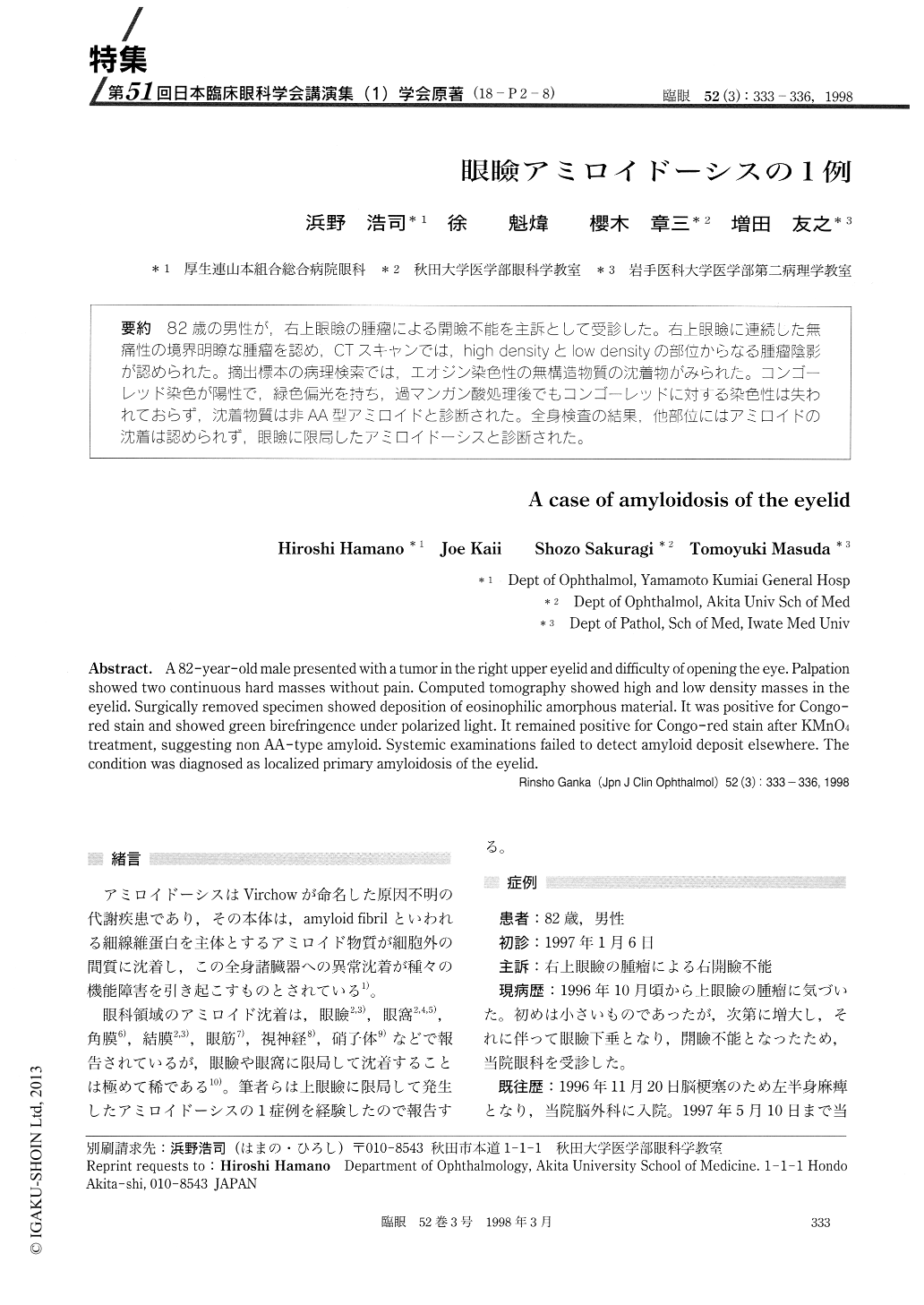
(18-P2-8) 82歳の男性が,右上眼瞼の腫瘤による開瞼不能を主訴として受診した。右上眼瞼に連続した無痛性の境界明瞭な腫瘤を認め,CTスキャンでは,high densityとlow densityの部位からなる腫瘤陰影が認められた。摘出標本の病理検索では,エオジン染色性の無構造物質の沈着物がみられた。コンゴーレッド染色が陽性で,緑色偏光を持ち,過マンガン酸処理後でもコンゴーレッドに対する染色性は失われておらず,沈着物質は非AA型アミロイドと診断された。全身検査の結果,他部位にはアミロイドの沈着は認められず,眼瞼に限局したアミロイドーシスと診断された。
A 82-year-old male presented with a tumor in the right upper eyelid and difficulty of opening the eye. Palpation showed two continuous hard masses without pain. Computed tomography showed high and low density masses in the eyelid. Surgically removed specimen showed deposition of eosinophilic amorphous material. It was positive for Congo-red stain and showed green birefringence under polarized light. It remained positive for Congo-red stain after KMnO4 treatment, suggesting non AA-type amyloid. Systemic examinations failed to detect amyloid deposit elsewhere. The condition was diagnosed as localized primary amyloidosis of the eyelid.

Copyright © 1998, Igaku-Shoin Ltd. All rights reserved.


