Japanese
English
- 有料閲覧
- Abstract 文献概要
- 1ページ目 Look Inside
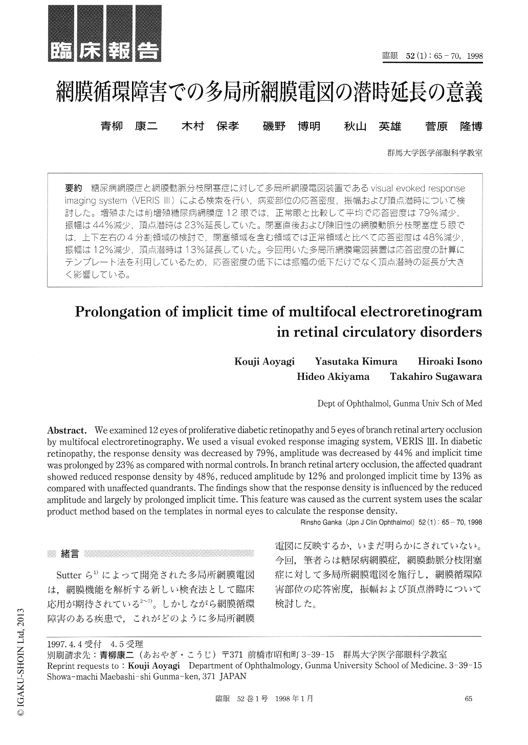
糖尿病網膜症と網膜動脈分枝閉塞症に対して多局所網膜電図装置であるvisual evoked responseimaging system (VERIS III)による検索を行い,病変部位の応答密度,振幅および頂点潜時について検討した。増殖または前増殖糖尿病網膜症12眼では,正常眼と比較して平均で応答密度は79%減少,振幅は44%減少,頂点潜時は23%延長していた。閉塞直後および陳旧性の網膜動脈分枝閉塞症5眼では,上下左右の4分割領域の検討で,閉塞領域を含む領域では正常領域と比べて応答密度は48%減少,振幅は12%減少,頂点潜時は13%延長していた。今回用いた多局所網膜電図装置は応答密度の計算にテンプレート法を利用しているため,応答密度の低下には振幅の低下だけでなく頂点潜時の延長が大きく影響している。
We examined 12 eyes of proliferative diabetic retinopathy and 5 eyes of branch retinal artery occlusion by multifocal electroretinography. We used a visual evoked response imaging system, VERIS III. In diabetic retinopathy, the response density was decreased by 79%, amplitude was decreased by 44% and implicit time was prolonged by 23% as compared with normal controls. In branch retinal artery occlusion, the affected quadrant showed reduced response density by 48%, reduced amplitude by 12% and prolonged implicit time by 13% as compared with unaffected quandrants. The findings show that the response density is influenced by the reduced amplitude and largely by prolonged implicit time. This feature was caused as the current system uses the scalar product method based on the templates in normal eyes to calculate the response density.

Copyright © 1998, Igaku-Shoin Ltd. All rights reserved.


