Japanese
English
- 有料閲覧
- Abstract 文献概要
- 1ページ目 Look Inside
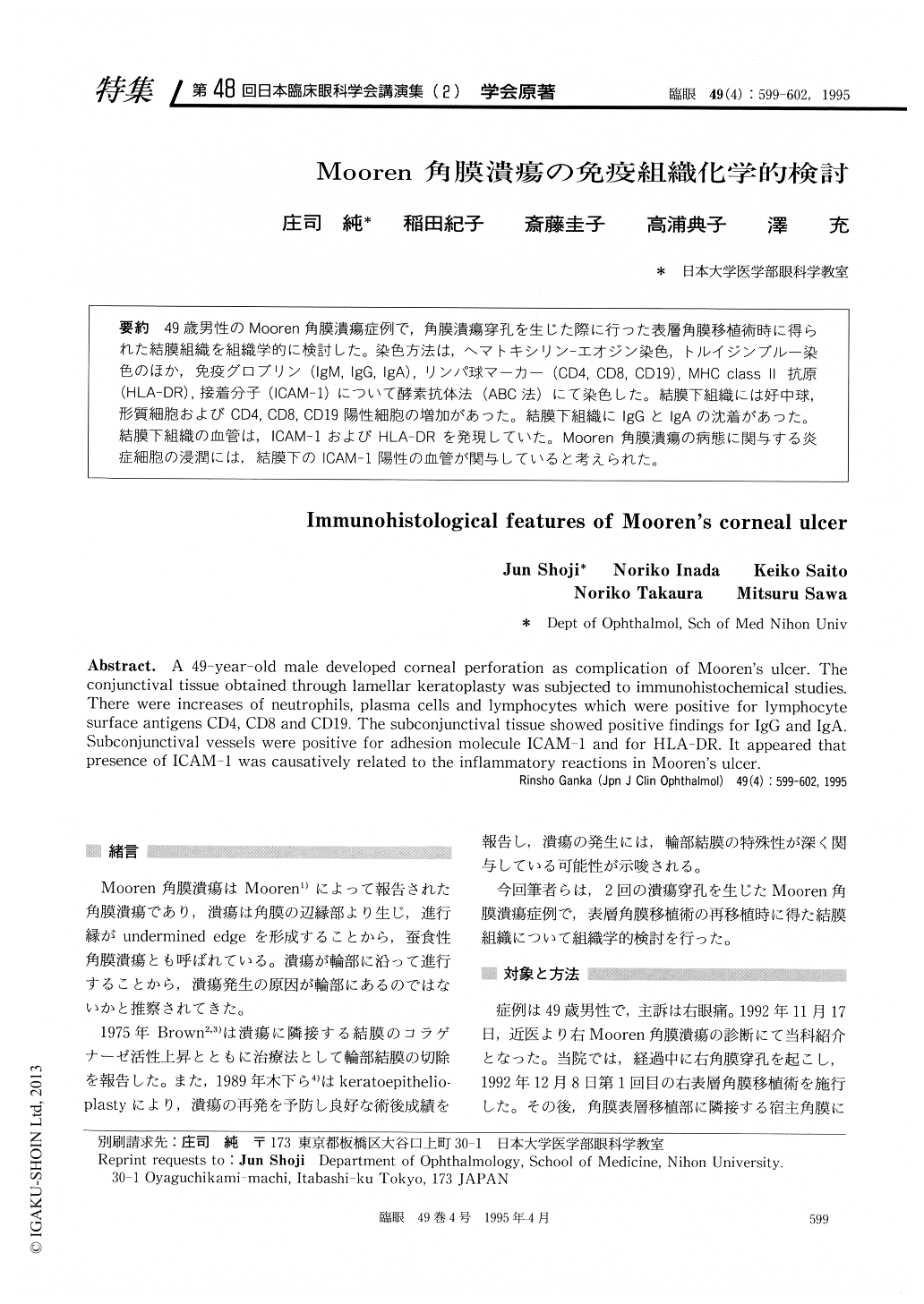
49歳男性のMooren角膜潰瘍症例で,角膜潰瘍穿孔を生じた際に行った表層角膜移植術時に得られた結膜組織を組織学的に検討した。染色方法は,ヘマトキシリン—エオジン染色,トルイジンブルー染色のほか,免疫グロブリン(IgM, IgG, IgA),リンパ球マーカー(CD4, CD8, CD19),MHC classⅡ抗原(HLA-DR),接着分子(ICAM−1)について酵素抗体法(ABC法)にて染色した。結膜下組織には好中球,形質細胞およびCD4,CD8,CD19陽性細胞の増加があった。結膜下組織にIgGとIgAの沈着があった。結膜下組織の血管は,ICAM−1およびHLA-DRを発現していた。Mooren角膜潰瘍の病態に関与する炎症細胞の浸潤には,結膜下のICAM−1陽性の血管が関与していると考えられた。
A 49-year-old male developed corneal perforation as complication of Mooren's ulcer. The conjunctival tissue obtained through lamellar keratoplasty was subjected to immunohistochemical studies. There were increases of neutrophils, plasma cells and lymphocytes which were positive for lymphocyte surface antigens CD4, CD8 and CD19. The subconjunctival tissue showed positive findings for IgG and IgA. Subconjunctival vessels were positive for adhesion molecule ICAM-1 and for HLA-DR. It appeared that presence of ICAM-1 was causatively related to the inflammatory reactions in Mooren's ulcer.

Copyright © 1995, Igaku-Shoin Ltd. All rights reserved.


