Japanese
English
- 有料閲覧
- Abstract 文献概要
- 1ページ目 Look Inside
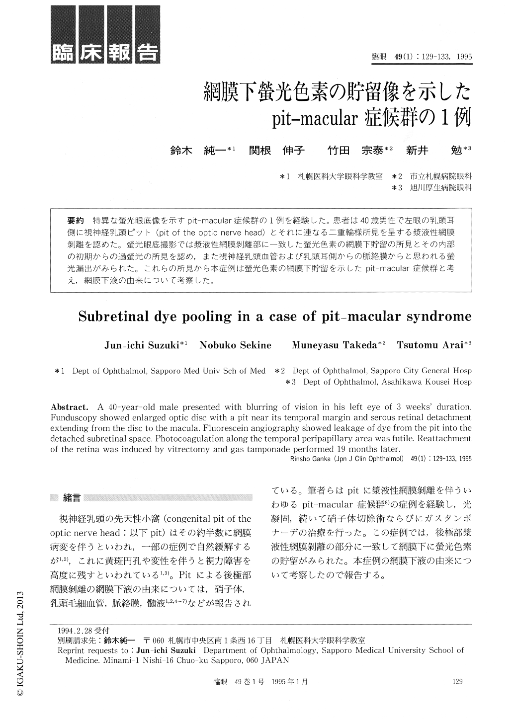
特異な螢光眼底像を示すpit-macular症候群の1例を経験した。患者は40歳男性で左眼の乳頭耳側に視神経乳頭ピット(pit of the optic nerve head)とそれに連なる二重輪様所見を呈する漿液性網膜剥離を認めた。螢光眼底撮影では漿液性網膜剥離部に一致した螢光色素の網膜下貯留の所見とその内部の初期からの過螢光の所見を認め,また視神経乳頭血管および乳頭耳側からの脈絡膜からと思われる螢光漏出がみられた。これらの所見から本症例は螢光色素の網膜下貯留を示したpit-macular症候群と考え,網膜下液の由来について考察した。
A 40-year-old male presented with blurring of vision in his left eye of 3 weeks' duration. Funduscopy showed enlarged optic disc with a pit near its temporal margin and serous retinal detachment extending from the disc to the macula. Fluorescein angiography showed leakage of dye from the pit into the detached subretinal space. Photocoagulation along the temporal peripapillary area was futile. Reattachment of the retina was induced by vitrectomy and gas tamponade performed 19 months later.

Copyright © 1995, Igaku-Shoin Ltd. All rights reserved.


