Japanese
English
- 有料閲覧
- Abstract 文献概要
- 1ページ目 Look Inside
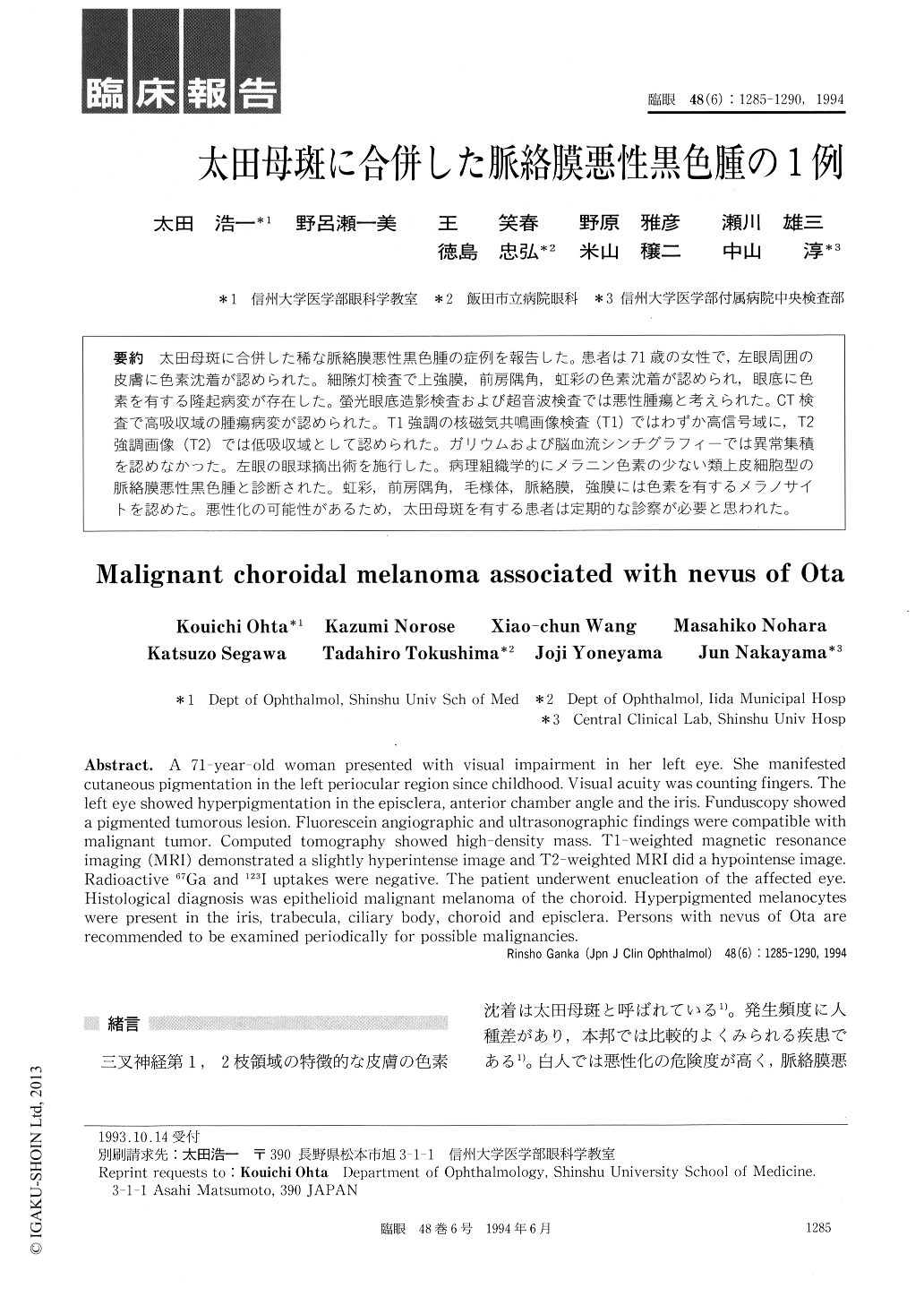
太田母斑に合併した稀な脈絡膜悪性黒色腫の症例を報告した。患者は71歳の女性で,左眼周囲の皮膚に色素沈着が認められた。細隙灯検査で上強膜,前房隅角,虹彩の色素沈着が認められ,眼底に色素を有する隆起病変が存在した。螢光眼底造影検査および超音波検査では悪性腫瘍と考えられた。CT検査で高吸収域の腫瘍病変が認められた。T1強調の核磁気共鳴画像検査(T1)ではわずか高信号域に,T2強調画像(T2)では低吸収域として認められた。ガリウムおよび脳血流シンチグラフィーでは異常集積を認めなかった。左眼の眼球摘出術を施行した。病理組織学的にメラニン色素の少ない類上皮細胞型の脈絡膜悪性黒色腫と診断された。虹彩,前房隅角,毛様体,脈絡膜,強膜には色素を有するメラノサイトを認めた。悪性化の可能性があるため,太田母斑を有する患者は定期的な診察が必要と思われた。
A 71-year-old woman presented with visual impairment in her left eye. She manifested cutaneous pigmentation in the left periocular region since childhood. Visual acuity was counting fingers. The left eye showed hyperpigmentation in the episclera, anterior chamber angle and the iris. Funduscopy showed a pigmented tumorous lesion. Fluorescein angiographic and ultrasonographic findings were compatible with malignant tumor. Computed tomography showed high-density mass. TI-weighted magnetic resonance imaging (MRI) demonstrated a slightly hyperintense image and T2-weighted MRI did a hypointense image. Radioactive 67Ga and 123I uptakes were negative. The patient underwent enucleation of the affected eye. Histological diagnosis was epithelioid malignant melanoma of the choroid. Hyperpigmented melanocytes were present in the iris, trabecula, ciliary body, choroid and episclera. Persons with nevus of Ota are recommended to be examined periodically for possible malignancies.

Copyright © 1994, Igaku-Shoin Ltd. All rights reserved.


