Japanese
English
- 有料閲覧
- Abstract 文献概要
- 1ページ目 Look Inside
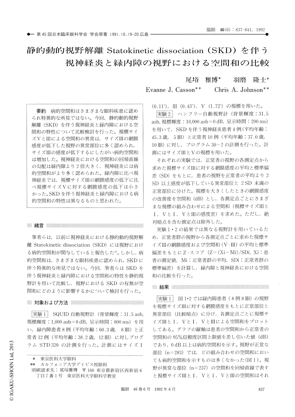
病的空間和はさまざまな眼科疾患に認められ特異的な所見ではない。今回,静的動的視野解離(SKD)を伴う視神経炎と緑内障における空間和の特性について比較検討を行った。視標サイズVとⅢによる空間和の異常は,サイズⅢの網膜感度が低下した視野の異常部位に多く認められ,サイズⅢの感度が低下するにしたがい病的空間和は増加した。視神経炎における空間和の回帰直線の勾配は緑内障より2倍大きく,視神経炎には病的空間和がより多く認められた。緑内障に比べ視神経炎では,視標サイズⅢの網膜感度の低下に比べ視標サイズVに対する網膜感度の低下は小さかった。SKDを伴う視神経炎と緑内障における病的空間和の特性は異なるものと思われた。
We tested the validity of our earlier observation that statokinetic dissociation in optic neuritis is the result of pathological spatial summation in the visual field. We compared the characteristics of spatial summation across the visual field in glaucoma and optic neuritis patients with statokinetic dissociation.
We performed automated static perimetry in 8 glaucoma eyes and 12 normal eyes. We used the Squid automated perimeter and compared the amount of spatial summation for various target sizes except absolute scotomas. The difference in sensitivity for size Ⅲ and V appeared to be mostappropriate for evaluation of spatial summation. We also used program 30-2 of Humphrey Field Analyzer in 5 optic neuritis patients and 10 normal eyes. Pathological spatial summation was observed in depressed areas in glaucoma and optic neuritis eyes. It was not obvious in normal visual field areas. The amount of spatial summation, or V minus Ⅲ, increased along with decrease in sensitiv ity to the size Ⅲ. The slope of regression line of Z-scores for spatial summation was twice as large in optic neuritis as in glaucoma. The sensitivity for size V at each eccentricity (0-10, 10-20, 20-30) was well spared in optic neuritis than in glaucoma. The findings suggest that the optic neuritis and glaucoma show different characteristics of pathological spatial summation.

Copyright © 1992, Igaku-Shoin Ltd. All rights reserved.


