Japanese
English
- 有料閲覧
- Abstract 文献概要
- 1ページ目 Look Inside
- 参考文献 Reference
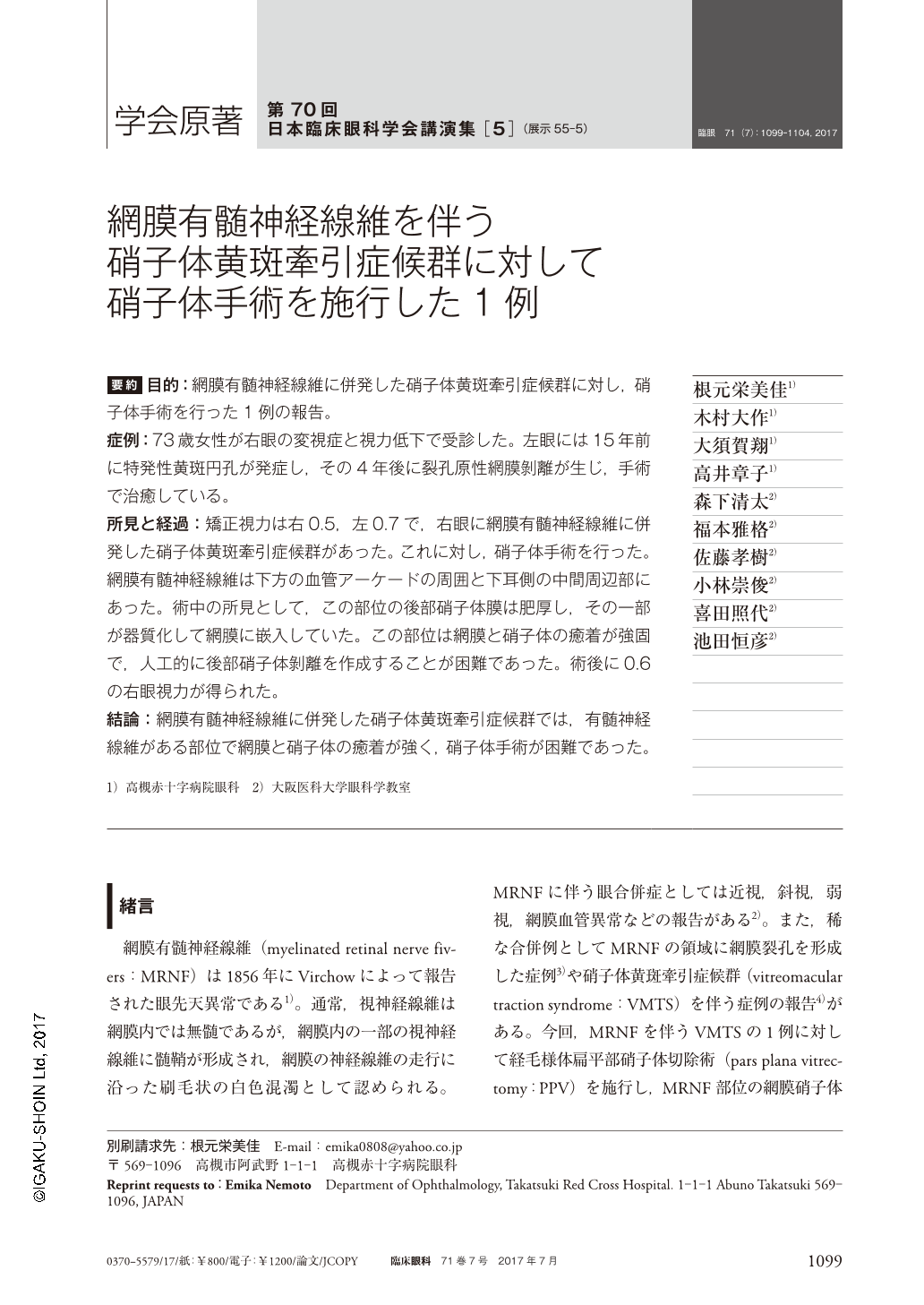
要約 目的:網膜有髄神経線維に併発した硝子体黄斑牽引症候群に対し,硝子体手術を行った1例の報告。
症例:73歳女性が右眼の変視症と視力低下で受診した。左眼には15年前に特発性黄斑円孔が発症し,その4年後に裂孔原性網膜剝離が生じ,手術で治癒している。
所見と経過:矯正視力は右0.5,左0.7で,右眼に網膜有髄神経線維に併発した硝子体黄斑牽引症候群があった。これに対し,硝子体手術を行った。網膜有髄神経線維は下方の血管アーケードの周囲と下耳側の中間周辺部にあった。術中の所見として,この部位の後部硝子体膜は肥厚し,その一部が器質化して網膜に嵌入していた。この部位は網膜と硝子体の癒着が強固で,人工的に後部硝子体剝離を作成することが困難であった。術後に0.6の右眼視力が得られた。
結論:網膜有髄神経線維に併発した硝子体黄斑牽引症候群では,有髄神経線維がある部位で網膜と硝子体の癒着が強く,硝子体手術が困難であった。
Abstract Purpose:To report a case of myelinated retinal nerve fiber with vitreous macular traction syndrome treated by vitreous surgery.
Case:A 73-year-old female presented with metamorphopsia and blurring in her right eye. She had developed idiopathic macular hole in the left eye 15 years before and rhegmatogenos retinal detachment 4 years later.
Findings and Clinical Course:Corrected visual acuity was 0.5 right and 0.7 left. The right eye showed myelinated retinal nerve fiber with vitreous macular traction syndrome. The right eye was treated by pars plana vitreous surgery. Myelinated retinal nerve fiber was present along the inferior vascular arcade and in the inferior temporal sector of midperipheral fundus. As intraoperative finding, the posterior vitreous facing the myelinated fiber showed thickening. A portion of thickened vitreous incarcerated into the retina. It was difficult to artificially detach the posterior vitreous from the retina. The right eye regained visual acuity of 0.6 after surgery.
Conclusion:The present case of myelinated retinal nerve fiber with vitreous macular traction syndrome presented difficulty in surgery due to adhesion of posterior vitreous to the site of myelinated retinal nerve fiber.

Copyright © 2017, Igaku-Shoin Ltd. All rights reserved.


