Japanese
English
- 有料閲覧
- Abstract 文献概要
- 1ページ目 Look Inside
- 参考文献 Reference
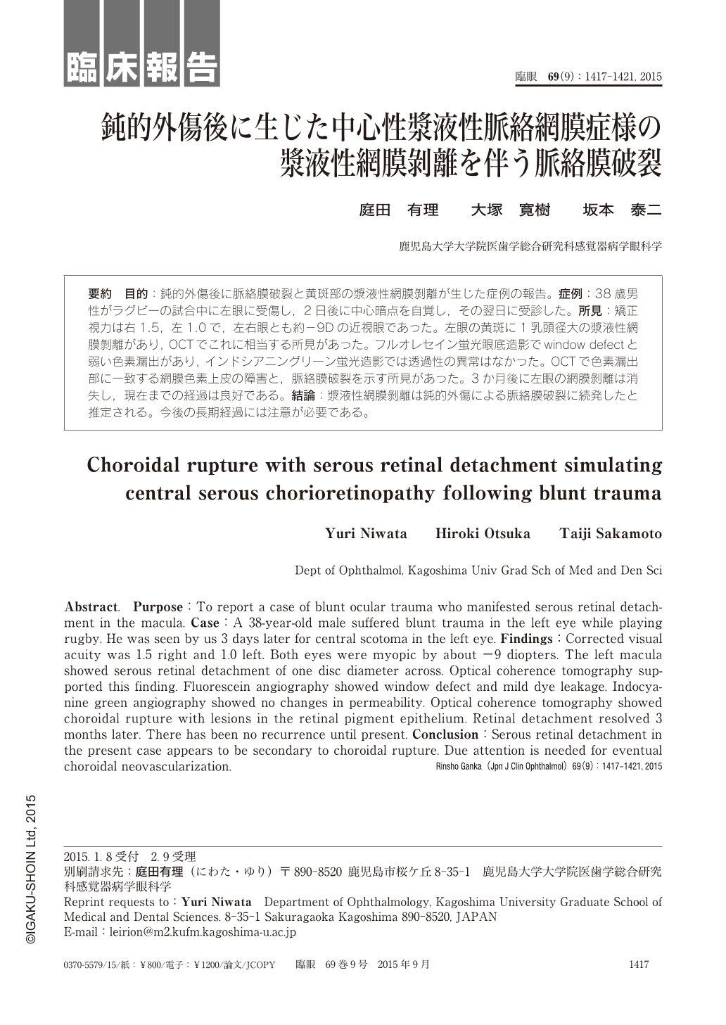
要約 目的:鈍的外傷後に脈絡膜破裂と黄斑部の漿液性網膜剝離が生じた症例の報告。症例:38歳男性がラグビーの試合中に左眼に受傷し,2日後に中心暗点を自覚し,その翌日に受診した。所見:矯正視力は右1.5,左1.0で,左右眼とも約−9Dの近視眼であった。左眼の黄斑に1乳頭径大の漿液性網膜剝離があり,OCTでこれに相当する所見があった。フルオレセイン蛍光眼底造影でwindow defectと弱い色素漏出があり,インドシアニングリーン蛍光造影では透過性の異常はなかった。OCTで色素漏出部に一致する網膜色素上皮の障害と,脈絡膜破裂を示す所見があった。3か月後に左眼の網膜剝離は消失し,現在までの経過は良好である。結論:漿液性網膜剝離は鈍的外傷による脈絡膜破裂に続発したと推定される。今後の長期経過には注意が必要である。
Abstract. Purpose:To report a case of blunt ocular trauma who manifested serous retinal detachment in the macula. Case:A 38-year-old male suffered blunt trauma in the left eye while playing rugby. He was seen by us 3 days later for central scotoma in the left eye. Findings:Corrected visual acuity was 1.5 right and 1.0 left. Both eyes were myopic by about −9 diopters. The left macula showed serous retinal detachment of one disc diameter across. Optical coherence tomography supported this finding. Fluorescein angiography showed window defect and mild dye leakage. Indocyanine green angiography showed no changes in permeability. Optical coherence tomography showed choroidal rupture with lesions in the retinal pigment epithelium. Retinal detachment resolved 3 months later. There has been no recurrence until present. Conclusion:Serous retinal detachment in the present case appears to be secondary to choroidal rupture. Due attention is needed for eventual choroidal neovascularization.

Copyright © 2015, Igaku-Shoin Ltd. All rights reserved.


