Japanese
English
- 有料閲覧
- Abstract 文献概要
- 1ページ目 Look Inside
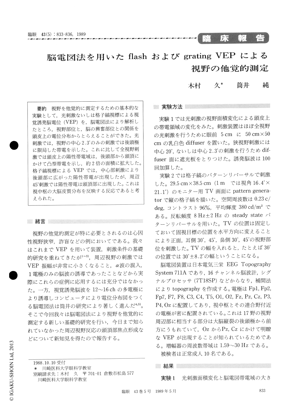
視野を他覚的に測定するための基本的な実験として,光刺激ないしは格子縞視標による視覚誘発脳電位(VEP)を,脳電図法により解析したところ,視野部位と,脳の興奮部位との関係を頭皮上の電位分布からとらえることができた。光刺激では,視野の中心2.3°のみの刺激では後頭極に限局した帯電を示した。これに比して全視野刺激では頭皮上の陽性帯電域は,後頭部から頭頂にかけて凸型帯電を示し,約2倍の面積に拡大した。格子縞視標によるVEPでは,中心部刺激により後頭部に広がった陽性帯電が出現したが,周辺45°刺激では陽性帯電は頭頂部に出現した。これは視中枢の大脳皮質分布を反映する反応であると考えられた。
We evaluated the topological correspondence between the stimulated area in the visual field and the excited cerebral cortex. We employed visually evoked cortical potential (VEP) elicited by flash or grating stimuli. The occipital area became positive-ly charged when the central visual field was stimulated by grating pattern. The parietal areaalso became positively charged when the peripheral visual field was stimulated in the same way. When the area of flash stimulus increased from the cen-tral 2.3° to the whole visual field, the positively charged area increased to twice the original. These results seemed to suggest that the VEP topography reflected functional locations in the cerebral cortex corresponding to the topography in the visual field.

Copyright © 1989, Igaku-Shoin Ltd. All rights reserved.


