Japanese
English
- 有料閲覧
- Abstract 文献概要
- 1ページ目 Look Inside
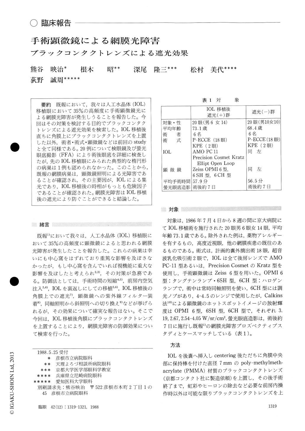
既報において,我々は人工水晶体(IOL)移植眼において35%の高頻度に手術顕微鏡光による網膜光障害が発生しうることを報告した.今回はその対策を検討する目的でブラックコンタクトレンズによる遮光効果を検索した.IOL移植後直ちに角膜上にブラックコンタクトレンズを上置した以外,術者・術式・顕微鏡などは前回のstudyと全て同様である.20例について検眼鏡及び螢光眼底撮影(FFA)により術後眼底を詳細に検査したが,先のIOL移植眼にみられた典型的な楕円形の病巣は1例も認められなかった.このことから,既報の網膜病巣は,顕微鏡照明による光障害であることが確認され,その主要因が,IOLによる集光であり,IOL移植後の時相がもっとも危険因子であることが確認された.網膜光障害はIOL移植後の遮光により防ぐことができると結論した.
In order to protect the retina from illumination by operating microscope during cataract surgery with intraocular lens implantation,we covered the cornea with black contact lens immediately after inserting the intraocular lens in the posterior cham-ber. Funduscopy was performed the next day and fluorescein fundus angiography one week after surgery.In a consecutive series of 20 eyes,it took an average of 27.4 minutes before extraction of nucleus, 15.7 minutes before lens insertion and 15.4 minutes before end of surgery,totalling 57.9 min-utes.
We observed no occurrence of photic retinopathy in the series, in sharp contract to the incidence of 35 % in a previous study by us under matching conditions concerning surgeons, surgical procedure, instrumentation and number of cases. Our findings show that our prior observations of retinal lesions were due to illumination through the implanted lens from the operating microscope. The photic retinal damage could completely be prevented by covering the cornea with a black contact lens immediately after insertion of intraocular lens.
Rinsho Ganka (Jpn J Clin Ophthalmol) 42(12):1319-1321, 1988

Copyright © 1988, Igaku-Shoin Ltd. All rights reserved.


