Japanese
English
- 有料閲覧
- Abstract 文献概要
- 1ページ目 Look Inside
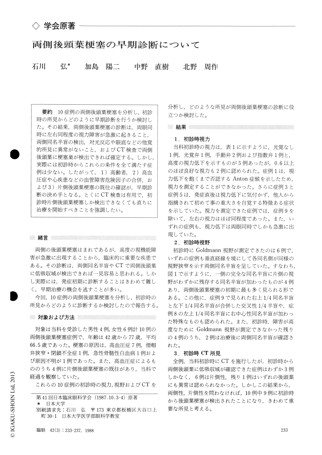
10症例の両側後頭葉梗塞を分析し,初診時の所見からどのように早期診断を行うか検討した.その結果,両側後頭葉梗塞の診断は,両眼同時に左右同程度の視力障害が急激に起きること,両側同名半盲の検出,対光反応や眼底などの他覚的所見に異常がないこと,およびCT検査で両側後頭葉に梗塞巣が検出できれば確定する.しかし,実際には初診時からこれらの条件を全て満たす症例は少ない.したがって,1)高齢者,2)高血圧症や心疾患などの血管障害危険因子の合併,および3)片側後頭葉梗塞の既往の確認が,早期診断の決め手となる.とくにCT検査は有用で,初診時片側後頭葉梗塞しか検出できなくても直ちに治療を開始すべきことを強調したい.
We evaluated a consecutive series of 10 cases with bilateral occipital infarction. The ages aver-age 66.5 years. Systemic hypertension was usually present. Four cases manifested a history of unilat-eral occipital infarction. Visual acuity was vari-able : 5 cases showed severe visual loss between no light perception and counting fingers, while 2 cases showed good visual acuity.
One case developed Anton's syndrome. Bilateral homonymous hemianopia was present in 6 cases. Presence of low density area in bilateral occipital lobes was detected in only 3 cases with CT scanningat the first examination. Still, CT scanning elicited the presence of occipital lobe infarction at least in one occipital lobe in 9 cases.
Our present series was characterized by sudden onset of visual loss, equal visual acuity for either eye, bilateral homonymous hemianopia, normal pupils and ocular fundus. Presence of low density areas in the occipital lobes by CT scanning was instrumental in the diagnosis.
Advanced age, generalized vascular lesions and past history of stroke, particularly unilateral oc-cipital infarction, were further features suggestive of bilateral involvement.
Rinsho Ganka (Jpn J Clin Ophthalmol) 42(3) : 233-237, 1988

Copyright © 1988, Igaku-Shoin Ltd. All rights reserved.


