Japanese
English
- 有料閲覧
- Abstract 文献概要
- 1ページ目 Look Inside
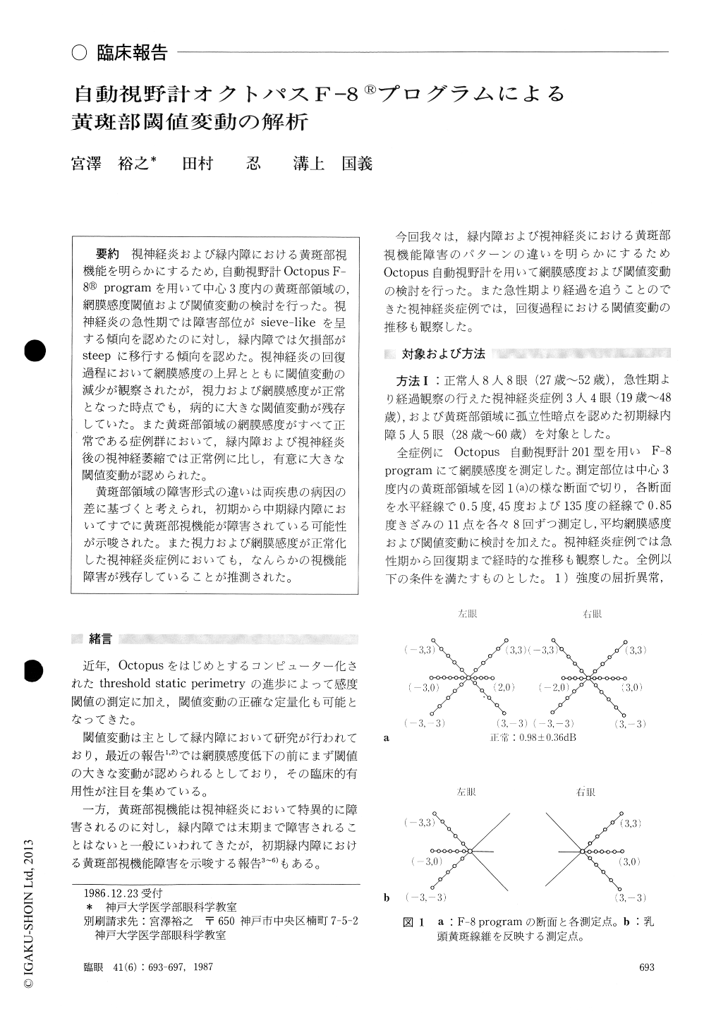
視神経炎および緑内障における黄斑部視機能を明らかにするため,自動視野計Octopus F-8®programを用いて中心3度内の黄斑部領域の,網膜感度閾値および閾値変動の検討を行った.視神経炎の急性期では障害部位がsieve-likeを呈する傾向を認めたのに対し,緑内障では欠損部がsteepに移行する傾向を認めた.視神経炎の回復過程において網膜感度の上昇とともに閾値変動の減少が観察されたが,視力および網膜感度が正常となった時点でも,病的に大きな閾値変動が残存していた.また黄斑部領域の網膜感度がすべて正常である症例群において,緑内障および視神経炎後の視神経萎縮では正常例に比し,有意に大きな閾値変動が認められた.
黄斑部領域の障害形式の違いは両疾患の病因の差に基づくと考えられ,初期から中期緑内障においてすでに黄斑部視機能が障害されている可能性が示唆された.また視力および網膜感度が正常化した視神経炎症例においても,なんらかの視機能障害が残存していることが推測された.
We measured the threshold and short-time fluctu-ations of foveal sensitivity in 8 normal subjects, 11 eyes in the convalescent period of optic neuritis, and 5 eyes with early glaucoma. We examined the profiles of the central, 3° visual field using programF-8 of Octopus 201 perimeter.
The short-term fluctuation was dependent on the retinal sensitivity. The fluctuation was larger when the sensitivity was low. Sieve-like depression was observed in optic neuritis in its acute phase. Isolated scotoma was observed in glaucoma. The slope of the scotoma was steeper in glaucoma than in optic neuritis.
The short-term flucutation gradually decreased during the course of recovery of optic neuritis. Still, wide short-time fluctuations persisted in spite of normalized retinal sensitivity in mean value. Theshort-time fluctuation was significantly wider than in patients with glaucoma or optic neuritis after apparent recovery than in normal subjects.
The findings indicate that macular function is disturbed in early to middle-stage glaucoma and after recovery of optic neuritis.
Rinsho Ganka (Jpn J Clin Ophthalmol) 41(6) : 693-697, 1987

Copyright © 1987, Igaku-Shoin Ltd. All rights reserved.


