Japanese
English
- 有料閲覧
- Abstract 文献概要
- 1ページ目 Look Inside
緒 言
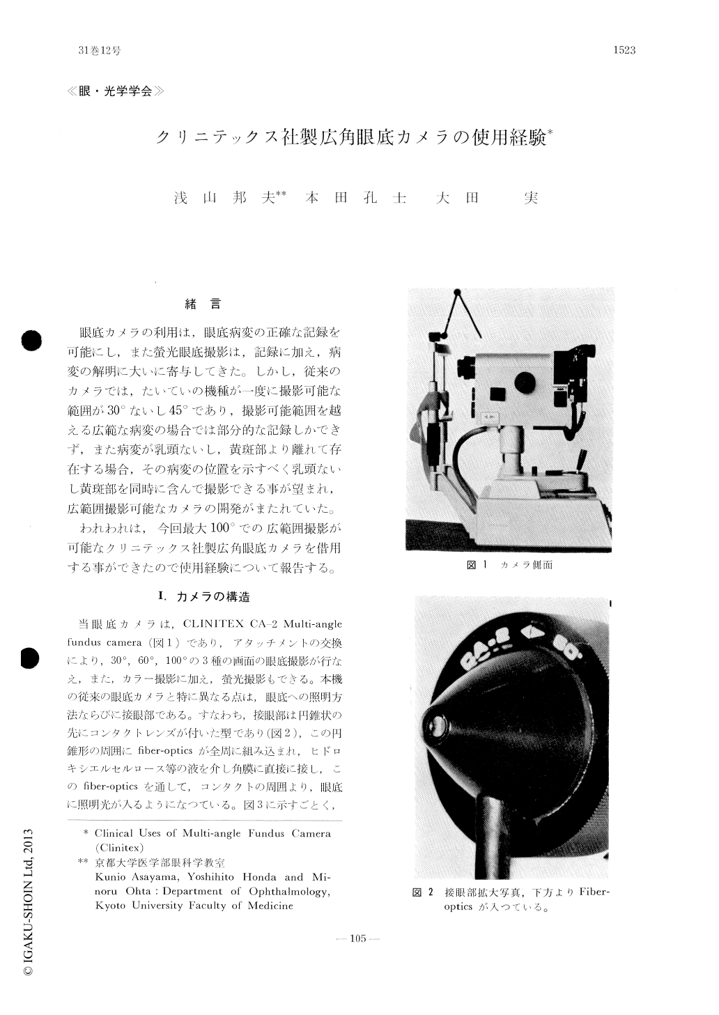
眼底カメラの利用は,眼底病変の正確な記録を可能にし,また螢光眼底撮影は,記録に加え,病変の解明に大いに寄与してきた。しかし,従来のカメラでは,たいていの機種が一度に撮影可能な範囲が30°ないし45°であり,撮影可能範囲を越える広範な病変の場合では部分的な記録しかできず,また病変が乳頭ないし,黄斑部より離れて存在する場合,その病変の位置を示すべく乳頭ないし黄斑部を同時に含んで撮影できる事が望まれ,広範囲撮影可能なカメラの開発がまたれていた。
われわれは,今回最大100°での広範囲撮影が可能なクリニテックス社製広角眼底カメラを借用する事ができたので使用経験について報告する。
We used a multi-angle fundus camera (Mo-del CA-2, Clinitex Inc.) for routine clinical stu-dies. Comments are made based on our clinical experiences. 1) We found it convenient that the operator can choose the angle of fundus photo-graphy from 30°, 60° and 100°. The quality of fundus pictures at 100° angle was gratifying and the pictures were very valuable for teaching purposes. 2) The manipulations of the camera was rather complicated and demanded an utmost cooperation of the examinee. 3) All the mechanics were set in the camera body. This feature facili-tated the manipulation in the dark.

Copyright © 1977, Igaku-Shoin Ltd. All rights reserved.


