Japanese
English
臨床実験
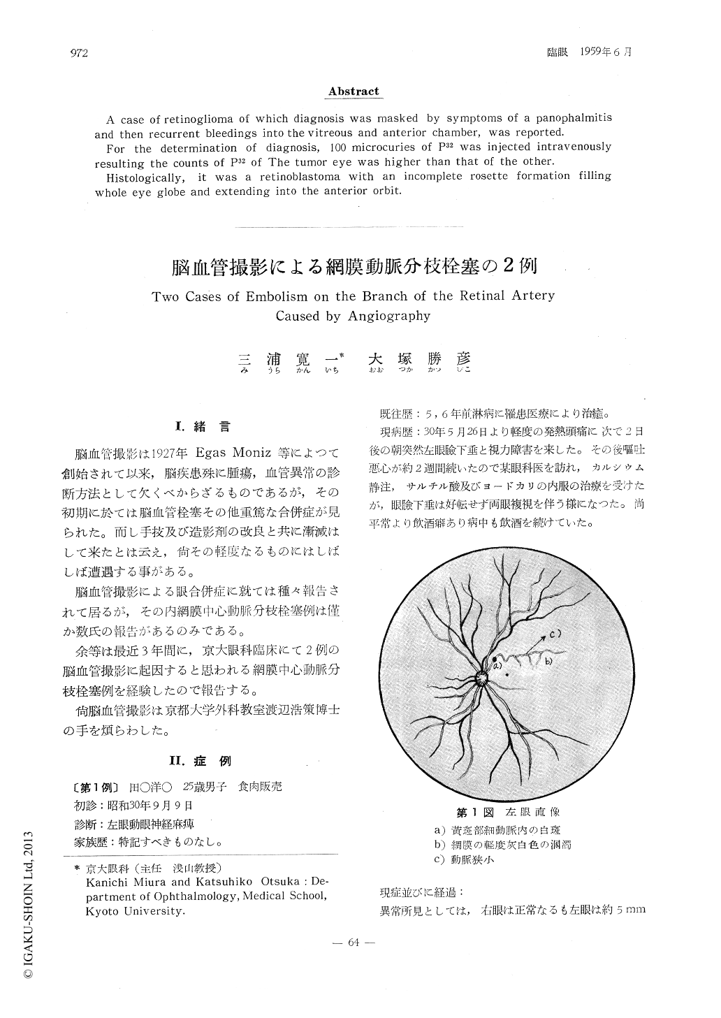
脳血管撮影による網膜動脈分枝栓塞の2例
Two Cases of Embolism on the Branch of the Retinal Artery Caused by Angiography
三浦 寛一
1
,
大塚 勝彦
1
Kanichi Miura
1
,
Katsuhiko Otsuka
1
1京大眼科
1Department of Ophthalmology, Medical School, Kyoto University.
pp.972-978
発行日 1959年6月15日
Published Date 1959/6/15
DOI https://doi.org/10.11477/mf.1410206693
- 有料閲覧
- Abstract 文献概要
- 1ページ目 Look Inside
I.緒言
脳血管撮影は1927年Egas Moniz等によつて創始されて以来,脳疾患殊に腫瘍,血管異常の診断方法として欠くべからざるものであるが,その初期に於ては脳血管栓塞その他重篤な合併症が見られた。而し手技及び造影剤の改良と共に漸減はして来たとは云え,尚その軽度なるものにはしばしば遭遇する事がある。
脳血管撮影による眼合併症に就ては種々報告されて居るが,その内網膜中心動脈分枝栓塞例は僅か数氏の報告があるのみである。
Two cases of embolism in the branch of the retinal artery possibly caused by cerebral angiography were reported.
Case 1. A 25 years old male developed embolism in the arteriole in the macular area of the left eye immediately after cerebral angiography with Uriodone.
Case 2. A 17 years old male developed embolism in the branch of temporal superior artery five days after cerebral angiography with Urografin.
Both cases showed defect of visual field without visual disturbance.

Copyright © 1959, Igaku-Shoin Ltd. All rights reserved.


