Japanese
English
- 有料閲覧
- Abstract 文献概要
- 1ページ目 Look Inside
緒言
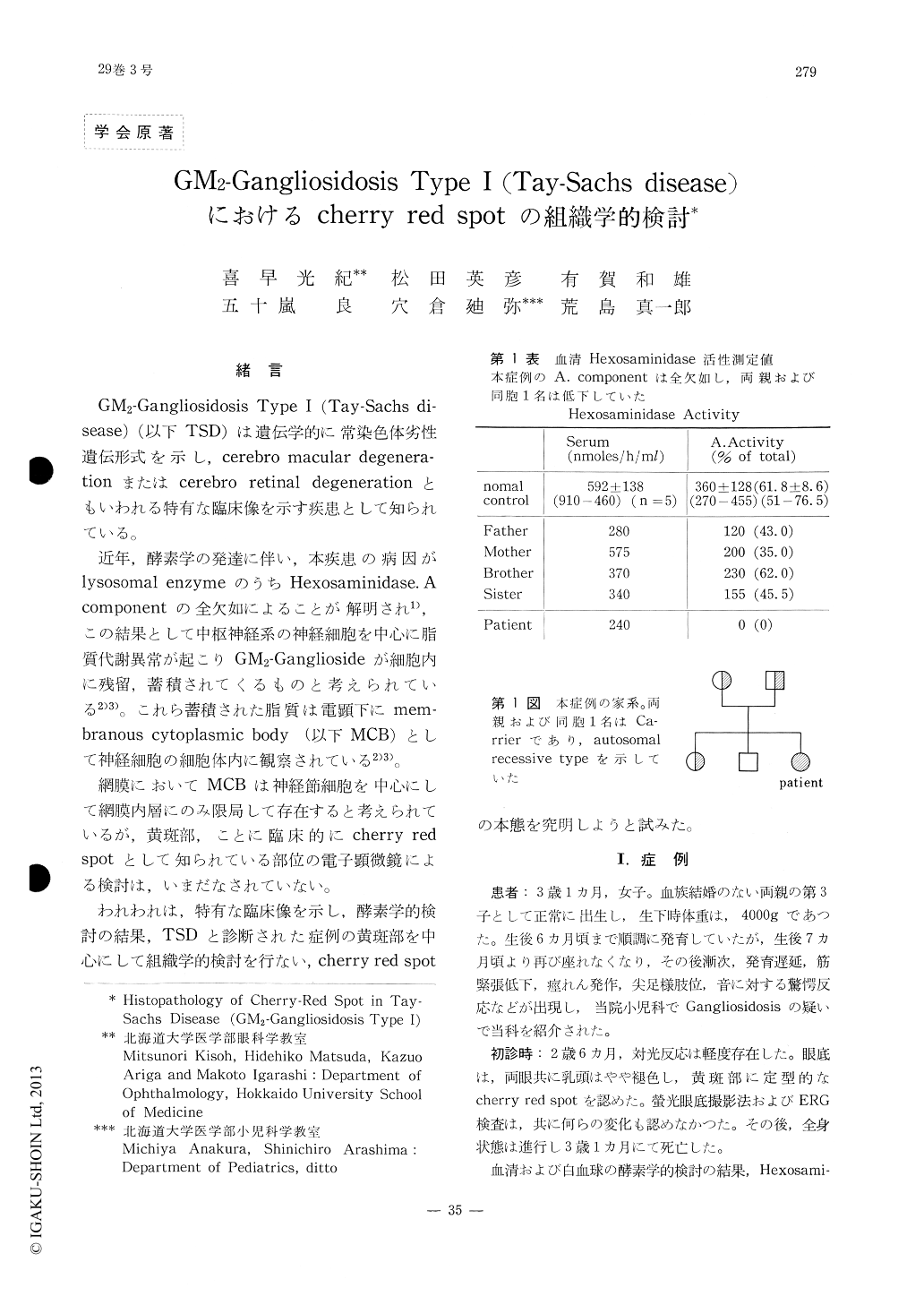
GM2—Gangliosidosis Type I (Tay-Sachs di—sease)(以下TSD)は遺伝学的に常染色体劣性遺伝形式を示し,cerebro macular degenera—tionまたはcerebro retinal degenerationともいわれる特有な臨床像を示す疾患として知られている。
近年,酵素学の発達に伴い,本疾患の病因がlysosomal enzymeのうちHexosaminidase.Acomponentの全欠如によることが解明され1),この結果として中枢神経系の神経細胞を中心に脂質代謝異常が起こりGM2—Gangliosideが細胞内に残留,蓄積されてくるものと考えられている2)3)。これら蓄積された脂質は電顕下にmem—branous cytoplasmic body (以下MCB)として神経細胞の細胞体内に観察されている2)3)。
The retina obtained from a case of Tay-Sa-chs disease was examined with both light and electron microscope to elucidate the pathogenesis of cherry-red spot.
At the area around the foveola, so called membranous cytoplasmic bodies were detected mainly in the ganglion cell layer. At the fo-veola, a few membranous cytoplasmic bodies were detected in the external nuclear layer and inner segments of photoreceptors. The diame-ter of the foveola measured in the specimen was consistent well with that of cherry-red spot calibrated in ocular fundus photograph.

Copyright © 1975, Igaku-Shoin Ltd. All rights reserved.


