Japanese
English
臨床実験
白点状網膜炎の螢光眼底所見
Fluorescein Angiography of Retinitis Punctata Albescens
五十嵐 良
1
,
田中 宣彦
1
Makoto Igarashi
1
,
Nobuhiko Tanaka
1
1国立札幌病院眼科
1Eye Clinic, Sapporo National Hospital
pp.2057-2061
発行日 1971年10月15日
Published Date 1971/10/15
DOI https://doi.org/10.11477/mf.1410204673
- 有料閲覧
- Abstract 文献概要
- 1ページ目 Look Inside
緒言
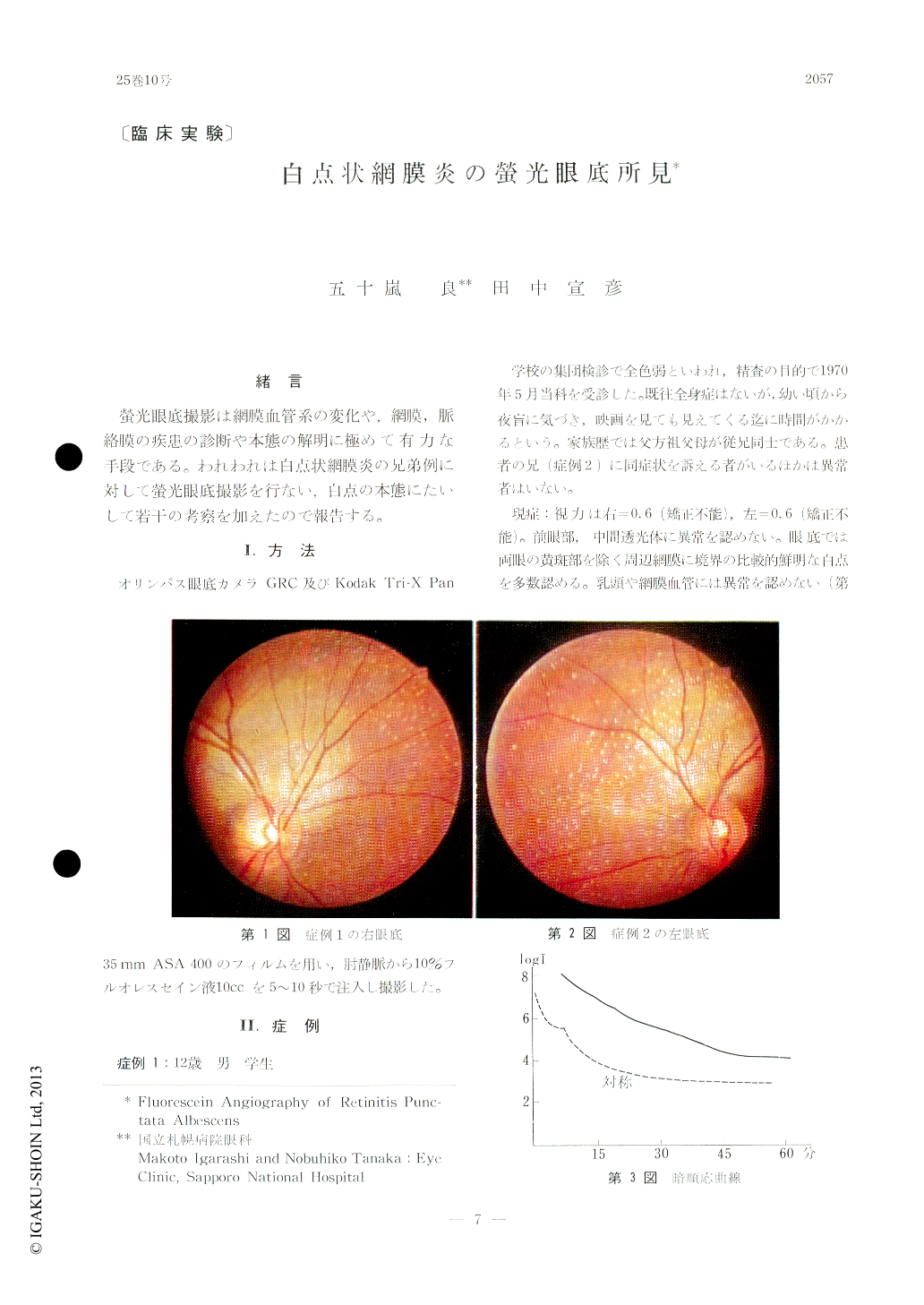
螢光眼底撮影は網膜血管系の変化や,網膜,脈絡膜の疾患の診断や本態の解明に極めて有力な手段である。われわれは白点状網膜炎の兄弟例に対して螢光眼底撮影を行ない,白点の本態にたいして若干の考察を加えたので報告する。
A report is presented over fluorescein fundus angiographic findings of progressive retinitis punctata albescens in two male siblings.
The characteristic white dots became distinct-ly fluorescent during the choroidal flush. After the arterial phase had set in, though, they be-came indiscernible due to multiple fluorescent foci which did not correspond to the white-dot lesions. These foci, or retinal dystrophic areas, showed neither leak or stain by fluorescein.

Copyright © 1971, Igaku-Shoin Ltd. All rights reserved.


