Japanese
English
- 有料閲覧
- Abstract 文献概要
- 1ページ目 Look Inside
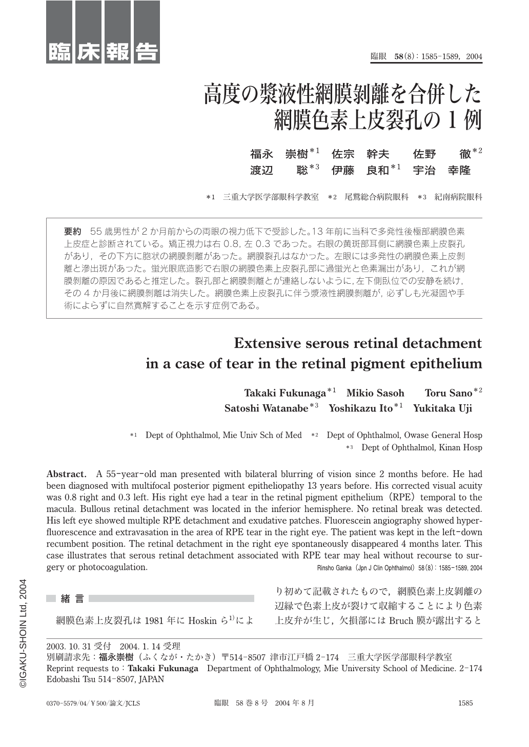
55歳男性が2か月前からの両眼の視力低下で受診した。13年前に当科で多発性後極部網膜色素上皮症と診断されている。矯正視力は右0.8,左0.3であった。右眼の黄斑部耳側に網膜色素上皮裂孔があり,その下方に胞状の網膜剝離があった。網膜裂孔はなかった。左眼には多発性の網膜色素上皮剝離と滲出斑があった。蛍光眼底造影で右眼の網膜色素上皮裂孔部に過蛍光と色素漏出があり,これが網膜剝離の原因であると推定した。裂孔部と網膜剝離とが連絡しないように,左下側臥位での安静を続け,その4か月後に網膜剝離は消失した。網膜色素上皮裂孔に伴う漿液性網膜剝離が,必ずしも光凝固や手術によらずに自然寛解することを示す症例である。
Abstract. A 55-year-old man presented with bilateral blurring of vision since 2months before. He had been diagnosed with multifocal posterior pigment epitheliopathy 13 years before. His corrected visual acuity was 0.8 right and 0.3 left. His right eye had a tear in the retinal pigment epithelium(RPE)temporal to the macula. Bullous retinal detachment was located in the inferior hemisphere. No retinal break was detected. His left eye showed multiple RPE detachment and exudative patches. Fluorescein angiography showed hyperfluorescence and extravasation in the area of RPE tear in the right eye. The patient was kept in the left-down recumbent position. The retinal detachment in the right eye spontaneously disappeared 4months later. This case illustrates that serous retinal detachment associated with RPE tear may heal without recourse to surgery or photocoagulation.

Copyright © 2004, Igaku-Shoin Ltd. All rights reserved.


