Japanese
English
- 有料閲覧
- Abstract 文献概要
- 1ページ目 Look Inside
- 参考文献 Reference
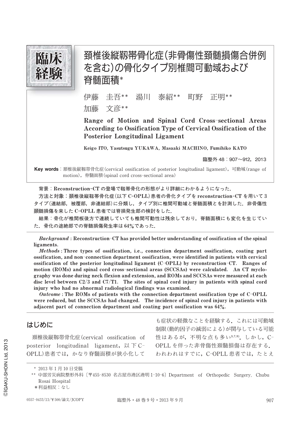
背景:Reconstruction-CTの登場で靱帯骨化の形態がより詳細にわかるようになった.
方法と対象:頚椎後縦靱帯骨化症(以下C-OPLL)患者の骨化タイプをreconstruction-CTを用いて3タイプ(連結部,被覆部,非連結部)に分類し,タイプ別に椎間可動域と脊髄面積とを計測した.非骨傷性頚髄損傷を来したC-OPLL患者では脊損発生部の検討をした.
結果:骨化が椎間板後方で連続していても椎間可動性は残余しており,脊髄面積にも変化を生じていた.骨化の途絶部での脊髄損傷発生率は64%であった.
Background:Reconstruction-CT has provided better understanding of ossification of the spinal ligaments.
Methods:Three types of ossification, i.e., connection department ossification, coating part ossification, and non-connection department ossification, were identified in patients with cervical ossification of the posterior longitudinal ligament (C-OPLL) by reconstruction-CT. Ranges of motion (ROMs) and spinal cord cross-sectional areas (SCCSAs) were calculated. An CT myclography was done during neck flexion and extension, and ROMs and SCCSAs were measured at each disc level between C2/3 and C7/T1. The sites of spinal cord injury in patients with spinal cord injury who had no abnormal radiological findings was examined.
Outcome:The ROMs of patients with the connection department ossification type of C-OPLL were reduced, but the SCCSAs had changed. The incidence of spinal cord injury in patients with adjacent part of connection department and coating part ossification was 64%.

Copyright © 2013, Igaku-Shoin Ltd. All rights reserved.


