Japanese
English
- 有料閲覧
- Abstract 文献概要
- 1ページ目 Look Inside
- 参考文献 Reference
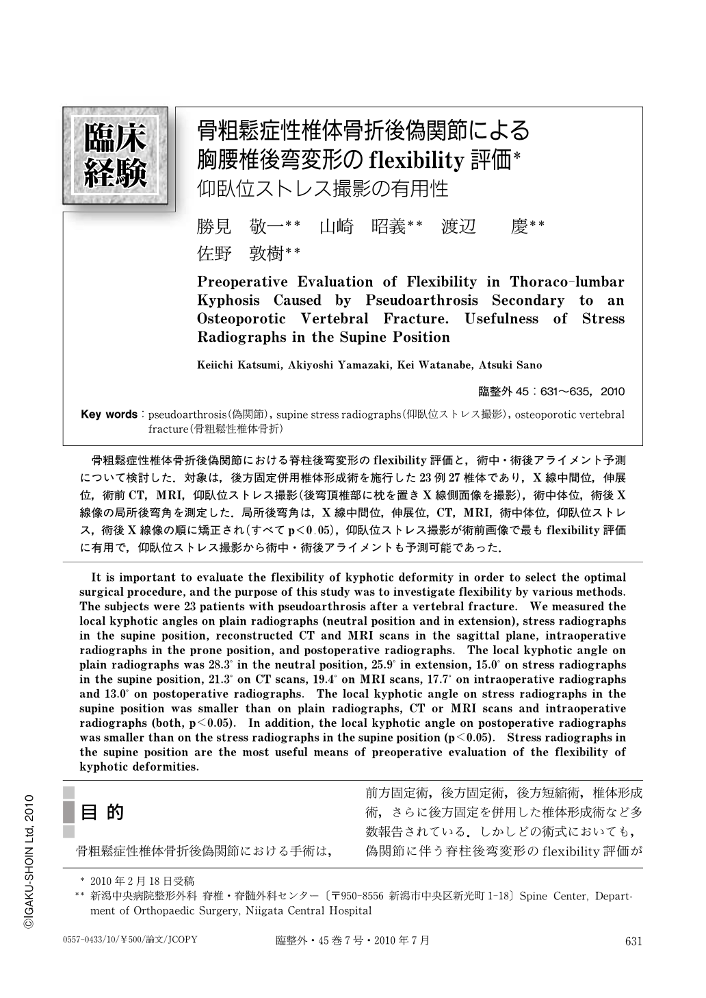
骨粗鬆症性椎体骨折後偽関節における脊柱後弯変形のflexibility評価と,術中・術後アライメント予測について検討した.対象は,後方固定併用椎体形成術を施行した23例27椎体であり,X線中間位,伸展位,術前CT,MRI,仰臥位ストレス撮影(後弯頂椎部に枕を置きX線側面像を撮影),術中体位,術後X線像の局所後弯角を測定した.局所後弯角は,X線中間位,伸展位,CT,MRI,術中体位,仰臥位ストレス,術後X線像の順に矯正され(すべてp<0.05),仰臥位ストレス撮影が術前画像で最もflexibility評価に有用で,仰臥位ストレス撮影から術中・術後アライメントも予測可能であった.
It is important to evaluate the flexibility of kyphotic deformity in order to select the optimal surgical procedure, and the purpose of this study was to investigate flexibility by various methods. The subjects were 23 patients with pseudoarthrosis after a vertebral fracture. We measured the local kyphotic angles on plain radiographs (neutral position and in extension), stress radiographs in the supine position, reconstructed CT and MRI scans in the sagittal plane, intraoperative radiographs in the prone position, and postoperative radiographs. The local kyphotic angle on plain radiographs was 28.3° in the neutral position, 25.9° in extension, 15.0° on stress radiographs in the supine position, 21.3° on CT scans, 19.4° on MRI scans, 17.7° on intraoperative radiographs and 13.0° on postoperative radiographs. The local kyphotic angle on stress radiographs in the supine position was smaller than on plain radiographs, CT or MRI scans and intraoperative radiographs (both, p<0.05). In addition, the local kyphotic angle on postoperative radiographs was smaller than on the stress radiographs in the supine position (p<0.05). Stress radiographs in the supine position are the most useful means of preoperative evaluation of the flexibility of kyphotic deformities.

Copyright © 2010, Igaku-Shoin Ltd. All rights reserved.


