Japanese
English
- 有料閲覧
- Abstract 文献概要
- 1ページ目 Look Inside
- 参考文献 Reference
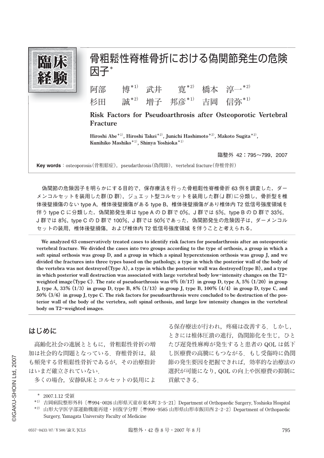
偽関節の危険因子を明らかにする目的で,保存療法を行った骨粗鬆性脊椎骨折63例を調査した.ダーメンコルセットを装用した群(D群),ジュエット型コルセットを装用した群(J群)に分類し,骨折型を椎体後壁損傷のないtype A,椎体後壁損傷があるtype B,椎体後壁損傷があり椎体内T2低信号強度領域を伴うtype Cに分類した.偽関節発生率はtype AのD群で0%,J群では5%,type BのD群で33%,J群では8%,type CのD群で100%,J群では50%であった.偽関節発生の危険因子は,ダーメンコルセットの装用,椎体後壁損傷,および椎体内T2低信号強度領域 を伴うことと考えられる.
We analyzed 63 conservatively treated cases to identify risk factors for pseudarthrosis after an osteoporotic vertebral fracture. We divided the cases into two groups according to the type of orthosis, a group in which a soft spinal orthosis was group D, and a group in which a spinal hyperextension orthosis was group J, and we divided the fractures into three types based on the pathology, a type in which the posterior wall of the body of the vertebra was not destroyed (Type A), a type in which the posterior wall was destroyed (type B), and a type in which posterior wall destruction was associated with large vertebral body low-intensity changes on the T2-weighted image (Type C). The rate of pseudoarthrosis was 0% (0/17) in group D, type A, 5% (1/20) in group J, type A, 33% (1/3) in group D, type B, 8% (1/13) in group J, type B, 100% (4/4) in group D, type C, and 50% (3/6) in group J, type C. The risk factors for pseudoarthrosis were concluded to be destruction of the posterior wall of the body of the vertebra, soft spinal orthosis, and large low intensity changes in the vertebral body on T2-weighted images.

Copyright © 2007, Igaku-Shoin Ltd. All rights reserved.


