Japanese
English
- 有料閲覧
- Abstract 文献概要
- 1ページ目 Look Inside
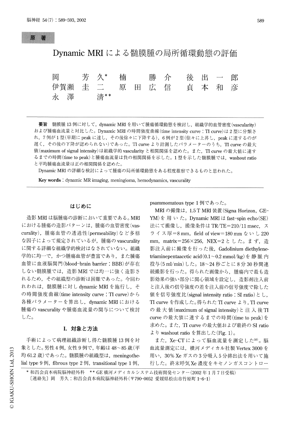
髄膜腫13例に対して,dynamic MRIを用いて腫瘍循環動態を検討し,組織学的血管密度(vascularity)および腫瘍血流量と対比した。Dynamic MRIの時間強度曲線(time intensity curve:TI curve)は2型に分類され,7例が1型(早期にpeakに達し,その後徐々に下降する),6例が2型(徐々に上昇し,peakに達するのが遅く,その後の下降が認められない)であった。TI curveより計測したパラメーターのうち,TI curveの最大値(maximum of signal intensity)は組織学的vascularityと相関関係を認めた。また,TI curveの最大値に達するまでの時間(time to peak)と腫瘍血流量は負の相関関係を示した。1型を示した髄膜腫では,washout ratioと平均腫瘍血流量は正の相関関係を認めた。
Dynamic MRIの詳細な検討によって腫瘍の局所循環動態をある程度推察できるものと思われた。
Dynamic MR imaging provides hemodynamic infor-mation about normal and pathologic tissue of the brain. The purpose of our study was to evaluate the usefulness of dynamic MR imaging in the assessment of tumor vascurality and the tumor tissue blood flow of meningiomas. We studied 13 patients with men-ingiomas using dynamic spin-echo MR imaging. The histological subtypes of meningioma were confirmed by the examination of surgical specimens in all pa-tients, and tumors were meningothelial in 9 cases, fi-brous in 2, transitional in 1, and psammomatous in 1.

Copyright © 2002, Igaku-Shoin Ltd. All rights reserved.


